Dominici M et al. (JAN 2006)
Cytotherapy 8 4 315--7
Minimal criteria for defining multipotent mesenchymal stromal cells. The International Society for Cellular Therapy position statement.
The considerable therapeutic potential of human multipotent mesenchymal stromal cells (MSC) has generated markedly increasing interest in a wide variety of biomedical disciplines. However,investigators report studies of MSC using different methods of isolation and expansion,and different approaches to characterizing the cells. Thus it is increasingly difficult to compare and contrast study outcomes,which hinders progress in the field. To begin to address this issue,the Mesenchymal and Tissue Stem Cell Committee of the International Society for Cellular Therapy proposes minimal criteria to define human MSC. First,MSC must be plastic-adherent when maintained in standard culture conditions. Second,MSC must express CD105,CD73 and CD90,and lack expression of CD45,CD34,CD14 or CD11b,CD79alpha or CD19 and HLA-DR surface molecules. Third,MSC must differentiate to osteoblasts,adipocytes and chondroblasts in vitro. While these criteria will probably require modification as new knowledge unfolds,we believe this minimal set of standard criteria will foster a more uniform characterization of MSC and facilitate the exchange of data among investigators.
View Publication
产品类型:
产品号#:
05420
05426
05429
05424
05434
产品名:
无动物成分的细胞解离试剂盒
Chute JP et al. (AUG 2006)
Proceedings of the National Academy of Sciences of the United States of America 103 31 11707--12
Inhibition of aldehyde dehydrogenase and retinoid signaling induces the expansion of human hematopoietic stem cells.
Aldehyde dehydrogenase (ALDH) is an enzyme that is expressed in the liver and is required for the conversion of retinol (vitamin A) to retinoic acids. ALDH is also highly enriched in hematopoietic stem cells (HSCs) and is considered a selectable marker of human HSCs,although its contribution to stem cell fate remains unknown. In this study,we demonstrate that ALDH is a key regulator of HSC differentiation. Inhibition of ALDH with diethylaminobenzaldehyde (DEAB) delayed the differentiation of human HSCs that otherwise occurred in response to cytokines. Moreover,short-term culture with DEAB caused a 3.4-fold expansion in the most primitive assayable human cells,the nonobese diabetic/severe combined immunodeficiency mouse repopulating cells,compared with day 0 CD34(+)CD38(-)lin(-) cells. The effects of DEAB on HSC differentiation could be reversed by the coadministration of the retinoic acid receptor agonist,all-trans-retinoic acid,suggesting that the ability of ALDH to generate retinoic acids is important in determining HSC fate. DEAB treatment also caused a decrease in retinoic acid receptor-mediated signaling within human HSCs,suggesting directly that inhibition of ALDH promotes HSC self-renewal via reduction of retinoic acid activity. Modulation of ALDH activity and retinoid signaling is a previously unrecognized and effective strategy to amplify human HSCs.
View Publication
产品类型:
产品号#:
01700
01705
01701
01702
产品名:
ALDEFLUOR™ 试剂盒
ALDEFLUOR™ DEAB试剂, 1.5 mM, 1 mL
ALDEFLUOR™检测缓冲液
Miyazaki T et al. (JAN 2014)
Genesis (New York,N.Y. : 2000) 52 1 49--55
Optimization of slow cooling cryopreservation for human pluripotent stem cells
Human pluripotent stem cells (hPSCs) have the potential for unlimited expansion and differentiation into cell types of all three germ layers. Cryopreservation is a key process for successful application of hPSCs. However,the current conventional method leads to poor recovery of hPSCs after thawing. Here,we demonstrate a highly efficient recovery method for hPSC cryopreservation by slow freezing and single-cell dissociation. After confirming hPSC survivability after freeze-thawing,we found that hPSCs that were freeze-thawed as colonies showed markedly decreased survival,whereas freeze-thawed single hPSCs retained the majority of their viability. These observations indicated that hPSCs should be cryopreserved as single cells. Freeze-thawed single hPSCs efficiently adhered and survived in the absence of a ROCK inhibitor by optimization of the seeding density. The high recovery rate enabled conventional colony passaging for subculture within 3 days post-thawing. The improved method was also adapted to a xeno-free culture system. Moreover,the cell recovery postcryopreservation was highly supported by coating culture surfaces with human laminin-521 that promotes adhesion of dissociated single hPSCs. This simplified but highly efficient cryopreservation method allows easy handling of cells and bulk storage of high-quality hPSCs.
View Publication
产品类型:
产品号#:
05860
05880
05850
05857
05870
05875
85850
85857
85870
85875
产品名:
mTeSR™1
mTeSR™1
Rapti K et al. (FEB 2015)
Molecular Therapy — Methods & Clinical Development 2 May 2014 14067
Effectiveness of gene delivery systems for pluripotent and differentiated cells.
Human embryonic stem cells (hESC) and induced pluripotent stem cells (hiPSC) assert a great future for the cardiovascular diseases,both to study them and to explore therapies. However,a comprehensive assessment of the viral vectors used to modify these cells is lacking. In this study,we aimed to compare the transduction efficiency of recombinant adeno-associated vectors (AAV),adenoviruses and lentiviral vectors in hESC,hiPSC,and the derived cardiomyocytes. In undifferentiated cells,adenoviral and lentiviral vectors were superior,whereas in differentiated cells AAV surpassed at least lentiviral vectors. We also tested four AAV serotypes,1,2,6,and 9,of which 2 and 6 were superior in their transduction efficiency. Interestingly,we observed that AAVs severely diminished the viability of undifferentiated cells,an effect mediated by induction of cell cycle arrest genes and apoptosis. Furthermore,we show that the transduction efficiency of the different viral vectors correlates with the abundance of their respective receptors. Finally,adenoviral delivery of the calcium-transporting ATPase SERCA2a to hESC and hiPSC-derived cardiomyocytes successfully resulted in faster calcium reuptake. In conclusion,adenoviral vectors prove to be efficient for both differentiated and undifferentiated lines,whereas lentiviral vectors are more applicable to undifferentiated cells and AAVs to differentiated cells.
View Publication
Lin P-Y et al. (NOV 2013)
Stem cells and development 23 4 372--379
A synthetic peptide-acrylate surface for production of insulin-producing cells from human embryonic stem cells.
Human embryonic stem cells (hESCs),due to their self-renewal capacity and pluripotency,have become a potential source of transplantable $\$-cells for the treatment of diabetes. However,it is imperative that the derived cells fulfill the criteria for clinical treatment. In this study,we replaced common Matrigel with a synthetic peptide-acrylate surface (Synthemax) to expand undifferentiated hESCs and direct their differentiation in a defined and serum-free medium. We confirmed that the cells still expressed pluripotent markers,had the ability to differentiate into three germ layers,and maintained a normal karyotype after 10 passages of subculture. Next,we reported an efficient protocol for deriving nearly 86% definitive endoderm cells from hESCs under serum-free conditions. Moreover,we were able to obtain insulin-producing cells within 21 days following a simple three-step protocol. The results of immunocytochemical and quantitative gene expression analysis showed that the efficiency of induction was not significantly different between the Synthemax surface and the Matrigel-coated surface. Thus,we provided a totally defined condition from hESC culture to insulin-producing cell differentiation,and the derived cells could be a therapeutic resource for diabetic patients in the future.
View Publication
产品类型:
产品号#:
05850
05857
05870
05875
85850
85857
85870
85875
产品名:
mTeSR™1
mTeSR™1
Roubal I et al. ( 2016)
Methods in molecular biology (Clifton,N.J.) 1341 345--357
Derivation of Neural Precursor Cells from Human Embryonic Stem Cells for DNA Methylomic Analysis.
Embryonic stem cells are self-renewing pluripotent cells with competency to differentiate into all three-germ lineages. Many studies have demonstrated the importance of genetic and epigenetic molecular mechanisms in the maintenance of self-renewal and pluripotency. Stem cells are under unique molecular and cellular regulations different from somatic cells. Proper regulation should be ensured to maintain their unique self-renewal and undifferentiated characteristics. Understanding key mechanisms in stem cell biology will be important for the successful application of stem cells for regenerative therapeutic medicine. More importantly practical use of stem cells will require our knowledge on how to properly direct and differentiate stem cells into the necessary type of cells. Embryonic stem cells and adult stem cells have been used as study models to unveil molecular and cellular mechanisms in various signaling pathways. They are especially beneficial to developmental studies where in vivo molecular/cellular study models are not available. We have derived neural stem cells from human embryonic stem cells as a model to study the effect of teratogen in neural development. We have tested commercial neural differentiation system and successfully derived neural precursor cells exhibiting key molecular features of neural stem cells,which will be useful for experimental application.
View Publication
产品类型:
产品号#:
05850
05857
05870
05875
85850
85857
85870
85875
产品名:
mTeSR™1
mTeSR™1
Haniffa M et al. (FEB 2009)
The Journal of experimental medicine 206 2 371--85
Differential rates of replacement of human dermal dendritic cells and macrophages during hematopoietic stem cell transplantation.
Animal models of hematopoietic stem cell transplantation have been used to analyze the turnover of bone marrow-derived cells and to demonstrate the critical role of recipient antigen-presenting cells (APC) in graft versus host disease (GVHD). In humans,the phenotype and lineage relationships of myeloid-derived tissue APC remain incompletely understood. It has also been proposed that the risk of acute GVHD,which extends over many months,is related to the protracted survival of certain recipient APC. Human dermis contains three principal subsets of CD45(+)HLA-DR(+) cells: CD1a(+)CD14(-) DC,CD1a(-)CD14(+) DC,and CD1a(-)CD14(+)FXIIIa(+) macrophages. In vitro,each subset has characteristic properties. After transplantation,both CD1a(+) and CD14(+) DC are rapidly depleted and replaced by donor cells,but recipient macrophages can be found in GVHD lesions and may persist for many months. Macrophages isolated from normal dermis secrete proinflammatory cytokines. Although they stimulate little proliferation of naive or memory CD4(+) T cells,macrophages induce cytokine expression in memory CD4(+) T cells and activation and proliferation of CD8(+) T cells. These observations suggest that dermal macrophages and DC are from distinct lineages and that persistent recipient macrophages,although unlikely to initiate alloreactivity,may contribute to GVHD by sustaining the responses of previously activated T cells.
View Publication
产品类型:
产品号#:
19155
19155RF
产品名:
Martin GR (DEC 1981)
Proceedings of the National Academy of Sciences of the United States of America 78 12 7634--8
Isolation of a pluripotent cell line from early mouse embryos cultured in medium conditioned by teratocarcinoma stem cells.
This report describes the establishment directly from normal preimplantation mouse embryos of a cell line that forms teratocarcinomas when injected into mice. The pluripotency of these embryonic stem cells was demonstrated conclusively by the observation that subclonal cultures,derived from isolated single cells,can differentiate into a wide variety of cell types. Such embryonic stem cells were isolated from inner cell masses of late blastocysts cultured in medium conditioned by an established teratocarcinoma stem cell line. This suggests that such conditioned medium might contain a growth factor that stimulates the proliferation or inhibits the differentiation of normal pluripotent embryonic cells,or both. This method of obtaining embryonic stem cells makes feasible the isolation of pluripotent cells lines from various types of noninbred embryo,including those carrying mutant genes. The availability of such cell lines should made possible new approaches to the study of early mammalian development.
View Publication
Gene expression profiling and localization of Hoechst-effluxing CD45- and CD45+ cells in the embryonic mouse lung.
Hoechst-effluxing cells (side population cells) are a rare subset of cells found in adult tissues that are highly enriched for stem and progenitor cell activity. To identify potential stem and progenitor cells during lung development,we generated gene expression profiles for CD45- and CD45+ side population cells in the embryonic day 17.5 lung. We found that side population cells comprise 1% of total embryonic day 17.5 lung cells (55% CD45+,45% CD45-). Gene profiling data demonstrated an overrepresentation of endothelial genes within the CD45- side population. We used expression of several distinct genes to identify two types of CD45- side population cells: 1) von Willebrand factor+/smooth muscle actin+ cells that reside in the muscular layer of select large vessels and 2) von Willebrand factor+/intercellular adhesion molecule+ cells that reside within the endothelial layer of select small vessels. Gene profiling of the CD45+ side population indicated an overrepresentation of genes associated with myeloid cell differentiation. Consistent with this,culturing CD45+ side population cells was associated with induction of mature dendritic markers (CD86). The microarray results suggested that expression of myeloperoxidase and proteinase-3 might be used to identify CD45+ side population cells. By immunohistochemistry,we found that myeloperoxidase+/proteinase-3+ cells represent a small subset of total CD45+ cells in the embryonic day 17.5 lung and that they reside in the mesenchyme and perivascular regions. This is the first detailed information regarding the phenotype and localization of side population cells in a developing organ.
View Publication
Siatskas C et al. (OCT 2005)
FASEB journal : official publication of the Federation of American Societies for Experimental Biology 19 12 1752--4
Specific pharmacological dimerization of KDR in lentivirally transduced human hematopoietic cells activates anti-apoptotic and proliferative mechanisms.
Selective and regulatable expansion of transduced cells could augment gene therapy for many disorders. The activation of modified growth factor receptors via synthetic chemical inducers of dimerization allows for the coordinated growth of transduced cells. This system can also provide information on specific receptor-mediated signaling without interference from other family members. Although several receptor subunits have been investigated in this context,little is known about the precise molecular events associated with dimerizer-initiated signaling. We have constructed and expressed an AP20187-regulated KDR chimeric receptor in human TF1 cells and analyzed activation of this gene switch using functional,biochemical,and microarray analyses. When deprived of natural ligands,GM-CSF,interleukin-3,or erythropoietin,AP20187 prevented apoptosis of transduced TF1 cells,induced dose-dependent proliferation,and supported long-term growth. In addition,AP20187 stimulation activated the signaling molecules associated with mitogen-activated protein kinase and phosphatidyl-inositol 3-kinase/Akt pathways. Microarray analysis determined that a number of transcripts involved in a variety of cellular processes were differentially expressed. Notably,mRNAs affiliated with heat stress,including Hsp70 and Hsp105,were up-regulated. Functional assays showed that Hsp70 and Hsp105 protected transduced TF1 cells from apoptosis and premature senescence,in part through regulation of Akt. These observations delineate specific roles for kinase insert domain-containing receptor,or KDR,signaling and suggest strategies to endow genetically modified cells with a survival advantage enabling the generation of adequate cell numbers for therapeutic outcomes.
View Publication

 EasySep™小鼠TIL(CD45)正选试剂盒


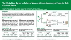 科学海报The Effect of Low Oxygen on Culture of Mouse and Human Mesenchymal Progenitor Cells from Bone Marrow

 沪公网安备31010102008431号