Haddad EA et al. (SEP 2009)
Journal of immunology (Baltimore,Md. : 1950) 183 6 3608--15
An accessory role for B cells in the IL-12-induced activation of resting mouse NK cells.
IL-12 is a potent proinflammatory cytokine. The effects of IL-12 are thought to be mediated by IFN-gamma production by NK,NKT,and T cells. In this study,we show that although IL-12 stimulates NK and NK1.1(+) T cells in bulk mouse splenocytes,it does not significantly stimulate purified NK cells,indicating that other cells are required. IL-12 stimulates T cell-deficient spleen cells and those depleted of macrophages. Unexpectedly,the depletion of dendritic cells also has little effect on the stimulation of spleen cells with IL-12. In contrast,B cell depletion almost completely inhibits IL-12-induced IFN-gamma production and B cell-deficient spleen cells are poorly stimulated with IL-12. Furthermore,purified NK cells are stimulated with IL-12 in the presence of purified B cells. Thus,B cells are necessary and also sufficient for the stimulation of purified NK cells with IL-12. Whereas spleen cells from IL-18-deficient mice are not stimulated with IL-12,NK cells purified from IL-18-deficient mice are stimulated with IL-12 in the presence of wild-type (WT) B cells,and WT NK cells are not stimulated with IL-12 in the presence of IL-18-deficient B cells. Cell contact between B and NK cells is also required for IL-12-induced IFN-gamma production. Finally,B cell-deficient mice injected with IL-12 produce significantly less IFN-gamma and IL-18 in the sera than WT mice do. Thus,stimulation of NK cells with IL-12 requires B cell cooperation in vitro as well as in vivo.
View Publication
产品类型:
产品号#:
18758
18758RF
18768
18768RF
产品名:
R. Lorenzetti et al. (jul 2019)
Journal of autoimmunity 101 145--152
Abatacept modulates CD80 and CD86 expression and memory formation in human B-cells.
BACKGROUND Cytotoxic T lymphocyte antigen-4 (CTLA-4) limits T-cell activation and is expressed on T-regulatory cells. Human CTLA-4 deficiency results in severe immune dysregulation. Abatacept (CTLA-4 Ig) is approved for the treatment of rheumatoid arthritis (RA) and its mechanism of action is attributed to effects on T-cells. It is known that CTLA-4 modulates the expression of its ligands CD80 and CD86 on antigen presenting cells (APC) by transendocytosis. As B-cells express CD80/CD86 and function as APC,we hypothesize that B-cells are a direct target of abatacept. OBJECTIVES To investigate direct effects of abatacept on human B-lymphocytes in vitro and in RA patients. METHODS The effect of abatacept on healthy donor B-cells' phenotype,activation and CD80/CD86 expression was studied in vitro. Nine abatacept-treated RA patients were studied. Seven of these were followed up to 24 months,and two up to 12 months only and treatment response,immunoglobulins,ACPA,RF concentrations,B-cell phenotype and ACPA-specific switched memory B-cell frequency were assessed. RESULTS B-cell development was unaffected by abatacept. Abatacept treatment resulted in a dose-dependent decrease of CD80/CD86 expression on B-cells in vitro,which was due to dynamin-dependent internalization. RA patients treated with abatacept showed a progressive decrease in plasmablasts and serum IgG. While ACPA-titers only moderately declined,the frequency of ACPA-specific switched memory B-cells significantly decreased. CONCLUSIONS Abatacept directly targets B-cells by reducing CD80/CD86 expression. Impairment of antigen presentation and T-cell activation may result in altered B-cell selection,providing a new therapeutic mechanism and a base for abatacept use in B-cell mediated autoimmunity.
View Publication
产品类型:
产品号#:
17954
17954RF
100-0971
产品名:
EasySep™人B细胞分选试剂盒
RoboSep™ 人B细胞分选试剂盒
EasySep™人B细胞分离试剂盒
Mace EM et al. ( 2016)
Nature communications 7 12171
Human NK cell development requires CD56-mediated motility and formation of the developmental synapse.
While distinct stages of natural killer (NK) cell development have been defined,the molecular interactions that shape human NK cell maturation are poorly understood. Here we define intercellular interactions between developing NK cells and stromal cells which,through contact-dependent mechanisms,promote the generation of mature,functional human NK cells from CD34(+) precursors. We show that developing NK cells undergo unique,developmental stage-specific sustained and transient interactions with developmentally supportive stromal cells,and that the relative motility of NK cells increases as they move through development in vitro and ex vivo. These interactions include the formation of a synapse between developing NK cells and stromal cells,which we term the developmental synapse. Finally,we identify a role for CD56 in developmental synapse structure,NK cell motility and NK cell development. Thus,we define the developmental synapse leading to human NK cell functional maturation.
View Publication
产品类型:
产品号#:
05150
15025
15065
产品名:
MyeloCult™ H5100
RosetteSep™人NK细胞富集抗体混合物
RosetteSep™人NK细胞富集抗体混合物
Garg TK et al. (SEP 2012)
Haematologica 97 9 1348--56
Highly activated and expanded natural killer cells for multiple myeloma immunotherapy.
BACKGROUND Patients with gene expression profiling-defined high-risk myeloma in relapse have poor outcomes with current therapies. We tested whether natural killer cells expanded by co-culture with K562 cells transfected with 41BBL and membrane-bound interleukin-15 could kill myeloma cells with a high-risk gene expression profile in vitro and in a unique model which recapitulates human myeloma. DESIGN AND METHODS OPM2 and high-risk primary myeloma tumors were grown in human fetal bone implanted into non-obese diabetic severe combined immunodeficiency mice with a deficient interleukin-2 receptor gamma chain. These mice are devoid of endogenous natural killer and T-cell activity and were used to determine whether adoptively transferred expanded natural killer cells could inhibit myeloma growth and myeloma-associated bone destruction. RESULTS Natural killer cells from healthy donors and myeloma patients expanded a median of 804- and 351-fold,respectively,without significant T-cell expansion. Expanded natural killer cells killed both allogeneic and autologous primary myeloma cells avidly via a perforin-mediated mechanism in which the activating receptor NKG2D,natural cytotoxicity receptors,and DNAX-accessory molecule-1 played a central role. Adoptive transfer of expanded natural killer cells inhibited the growth of established OPM2 and high-risk primary myeloma tumors grown in the murine model. The transferred,expanded natural killer cells proliferated in vivo in an interleukin-2 dose-dependent fashion,persisted up to 4 weeks,were readily detectable in the human bone,inhibited myeloma growth and protected bone from myeloma-induced osteolysis. CONCLUSIONS These studies provide the rationale for testing expanded natural killer cells in humans.
View Publication
产品类型:
产品号#:
19055
19055RF
产品名:
EasySep™人NK细胞富集试剂盒
RoboSep™ 人NK细胞富集试剂盒含滤芯吸头
A. Lopresti et al. (jun 2019)
JCI insight 5
Sensitive and easy screening for circulating tumor cells by flow cytometry.
Circulating Tumor Cells (CTCs) represent an easy,repeatable and representative access to information regarding solid tumors. However,their detection remains difficult because of their paucity,their short half-life,and the lack of reliable surface biomarkers. Flow cytometry (FC) is a fast,sensitive and affordable technique,ideal for rare cells detection. Adapted to CTCs detection (i.e. extremely rare cells),most FC-based techniques require a time-consuming pre-enrichment step,followed by a 2-hours staining procedure,impeding on the efficiency of CTCs detection. We overcame these caveats and reduced the procedure to less than one hour,with minimal manipulation. First,cells were simultaneously fixed,permeabilized,then stained. Second,using low-speed FC acquisition conditions and two discriminators (cell size and pan-cytokeratin expression),we suppressed the pre-enrichment step. Applied to blood from donors with or without known malignant diseases,this protocol ensures a high recovery of the cells of interest independently of their epithelial-mesenchymal plasticity and can predict which samples are derived from cancer donors. This proof-of-concept study lays the bases of a sensitive tool to detect CTCs from a small amount of blood upstream of in-depth analyses.
View Publication
Tyznik AJ et al. ( 2014)
The Journal of Immunology 192 8 3676--85
Distinct requirements for activation of NKT and NK cells during viral infection
NK cells are key regulators of innate defense against mouse CMV (MCMV). Like NK cells,NKT cells also produce high levels of IFN-γ rapidly after MCMV infection. However,whether similar mechanisms govern activation of these two cell types,as well as the significance of NKT cells for host resistance,remain unknown. In this article,we show that,although both NKT and NK cells are activated via cytokines,their particular cytokine requirements differ significantly in vitro and in vivo. IL-12 is required for NKT cell activation in vitro but is not sufficient,whereas NK cells have the capacity to be activated more promiscuously in response to individual cytokines from innate cells. In line with these results,GM-CSF-derived dendritic cells activated only NK cells upon MCMV infection,consistent with their virtual lack of IL-12 production,whereas Flt3 ligand-derived dendritic cells produced IL-12 and activated both NK and NKT cells. In vivo,NKT cell activation was abolished in IL-12(-/-) mice infected with MCMV,whereas NK cells were still activated. In turn,splenic NK cell activation was more IL-18 dependent. The differential requirements for IL-12 and IL-18 correlated with the levels of cytokine receptor expression by NK and NKT cells. Finally,mice lacking NKT cells showed reduced control of MCMV,and depleting NK cells further enhanced viral replication. Taken together,our results show that NKT and NK cells have differing requirements for cytokine-mediated activation,and both can contribute nonredundantly to MCMV defense,revealing that these two innate lymphocyte subsets function together to fine-tune antiviral responses.
View Publication
产品类型:
产品号#:
21000
20119
20155
18554
18554RF
18564
18564RF
产品名:
RoboSep™- S
RoboSep™ 吸头组件抛光剂
RoboSep™分选管套装(9个塑料管)
Benvenuto F et al. (JUL 2007)
Stem cells (Dayton,Ohio) 25 7 1753--60
Human mesenchymal stem cells promote survival of T cells in a quiescent state.
Mesenchymal stem cells (MSC) are part of the bone marrow that provides signals supporting survival and growth of bystander hematopoietic stem cells (HSC). MSC modulate also the immune response,as they inhibit proliferation of lymphocytes. In order to investigate whether MSC can support survival of T cells,we investigated MSC capacity of rescuing T lymphocytes from cell death induced by different mechanisms. We observed that MSC prolong survival of unstimulated T cells and apoptosis-prone thymocytes cultured under starving conditions. MSC rescued T cells from activation induced cell death (AICD) by downregulation of Fas receptor and Fas ligand on T cell surface and inhibition of endogenous proteases involved in cell death. MSC dampened also Fas receptor mediated apoptosis of CD95 expressing Jurkat leukemic T cells. In contrast,rescue from AICD was not associated with a significant change of Bcl-2,an inhibitor of apoptosis induced by cell stress. Accordingly,MSC exhibited a minimal capacity of rescuing Jurkat cells from chemically induced apoptosis,a process disrupting the mitochondrial membrane potential regulated by Bcl-2. These results suggest that MSC interfere with the Fas receptor regulated process of programmed cell death. Overall,MSC can inhibit proliferation of activated T cells while supporting their survival in a quiescent state,providing a model of their activity inside the HSC niche. Disclosure of potential conflicts of interest is found at the end of this article.
View Publication
产品类型:
产品号#:
05401
05402
05411
产品名:
MesenCult™ MSC 基础培养基(人)
MesenCult™ MSC 刺激补充剂(人)
MesenCult™ 增殖试剂盒(人)
C. Onyilagha et al. (jun 2019)
Journal of immunology (Baltimore,Md. : 1950)
NK Cells Are Critical for Optimal Immunity to Experimental Trypanosoma congolense Infection.
NK cells are key innate immune cells that play critical roles in host defense. Although NK cells have been shown to regulate immunity to some infectious diseases,their role in immunity to Trypanosoma congolense has not been investigated. NK cells are vital sources of IFN-gamma and TNF-alpha; two key cytokines that are known to play important roles in resistance to African trypanosomes. In this article,we show that infection with T. congolense leads to increased levels of activated and functional NK cells in multiple tissue compartments. Systemic depletion of NK cells with anti-NK1.1 mAb led to increased parasitemia,which was accompanied by significant reduction in IFN-gamma production by immune cells in the spleens and liver of infected mice. Strikingly,infected NFIL3-/- mice (which genetically lack NK cell development and function) on the normally resistant background were highly susceptible to T. congolense infection. These mice developed fulminating and uncontrolled parasitemia and died significantly earlier (13 ± 1 d) than their wild-type control mice (106 ± 26 d). The enhanced susceptibility of NFIL3-/- mice to infection was accompanied by significantly impaired cytokine (IFN-gamma and TNF-alpha) response by CD3+ T cells in the spleens and liver. Adoptive transfer of NK cells into NFIL3-/- mice before infection rescued them from acute death in a perforin-dependent manner. Collectively,these studies show that NK cells are critical for optimal resistance to T. congolense,and its deficiency leads to enhanced susceptibility in infected mice.
View Publication
产品类型:
产品号#:
19855
19855RF
产品名:
EasySep™小鼠NK细胞分选试剂盒
RoboSep™ 小鼠NK细胞分选试剂盒
Summers-DeLuca LE et al. (MAY 2007)
The Journal of experimental medicine 204 5 1071--81
Expression of lymphotoxin-alphabeta on antigen-specific T cells is required for DC function.
During an immune response,activated antigen (Ag)-specific T cells condition dendritic cells (DCs) to enhance DC function and survival within the inflamed draining lymph node (LN). It has been difficult to ascertain the role of the tumor necrosis factor (TNF) superfamily member lymphotoxin-alphabeta (LTalphabeta) in this process because signaling through the LTbeta-receptor (LTbetaR) controls multiple aspects of lymphoid tissue organization. To resolve this,we have used an in vivo system where the expression of TNF family ligands is manipulated only on the Ag-specific T cells that interact with and condition Ag-bearing DCs. We report that LTalphabeta is a critical participant required for optimal DC function,independent of its described role in maintaining lymphoid tissue organization. In the absence of LTalphabeta or CD40L on Ag-specific T cells,DC dysfunction could be rescued in vivo via CD40 or LTbetaR stimulation,respectively,suggesting that these two pathways cooperate for optimal DC conditioning.
View Publication

 EasySep™小鼠TIL(CD45)正选试剂盒


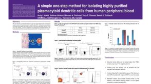 科学海报Isolation of Plasmacytoid Dendritic Cells from Human Peripheral Blood
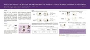 科学海报Pre-Enrichment of Dendritic Cells from Human Peripheral Blood Samples

 沪公网安备31010102008431号