Identification of rare HIV-1-infected patients with extreme CD4+ T cell decline despite ART-mediated viral suppression.
BACKGROUND The goal of antiretroviral therapy (ART) is to suppress HIV-1 replication and reconstitute CD4+ T cells. Here,we report on HIV-infected individuals who had a paradoxical decline in CD4+ T cells despite ART-mediated suppression of plasma HIV-1 load (pVL). We defined such an immunological outcome as extreme immune decline (EXID). METHODS EXID's clinical and immunological characteristics were compared to immunological responders (IRs),immunological nonresponders (INRs),healthy controls (HCs),and idiopathic CD4+ lymphopenia (ICL) patients. T cell immunophenotyping and assembly/activation of inflammasomes were evaluated by flow cytometry. PBMC transcriptome analysis and genetic screening for pathogenic variants were performed. Levels of cytokines/chemokines were measured by electrochemiluminescence. Luciferase immunoprecipitation system and NK-mediated antibody-dependent cellular cytotoxicity (ADCC) assays were used to identify anti-lymphocyte autoantibodies. RESULTS EXIDs were infected with non-B HIV-1 subtypes and after 192 weeks of consistent ART-mediated pVL suppression had a median CD4+ decrease of 157 cells/mul,compared with CD4+ increases of 193 cells/mul and 427 cells/mul in INR and IR,respectively. EXID had reduced naive CD4+ T cells,but similar proportions of cycling CD4+ T cells and HLA-DR+CD38+CD8+ T cells compared with IR and INR. Levels of inflammatory cytokines were also similar in EXID and INR,but the IL-7 axis was profoundly perturbed compared with HC,IR,INR,and ICL. Genes involved in T cell and monocyte/macrophage function,autophagy,and cell migration were differentially expressed in EXID. Two of the 5 EXIDs had autoantibodies causing ADCC,while 2 different EXIDs had an increased inflammasome/caspase-1 activation despite consistently ART-suppressed pVL. CONCLUSIONS EXID is a distinct immunological outcome compared with previously described INR. Anti-CD4+ T cell autoantibodies and aberrant inflammasome/caspase-1 activation despite suppressed HIV-1 viremia are among the mechanisms responsible for EXID.
View Publication
产品类型:
产品号#:
17955
17955RF
100-0960
产品名:
EasySep™人NK细胞分选试剂盒
RoboSep™ 人NK细胞分选试剂盒
EasySep™人NK细胞分离试剂盒
D. M. Previte et al. (apr 2019)
Cell reports 27 1 129--141.e4
Lymphocyte Activation Gene-3 Maintains Mitochondrial and Metabolic Quiescence in Naive CD4+ T Cells.
Lymphocyte activation gene-3 (LAG-3) is an inhibitory receptor expressed by CD4+ T cells and tempers their homeostatic expansion. Because CD4+ T cell proliferation is tightly coupled to bioenergetics,we investigate the role of LAG-3 in modulating naive CD4+ T cell metabolism. LAG-3 deficiency enhances the metabolic profile of naive CD4+ T cells by elevating levels of mitochondrial biogenesis. In vivo,LAG-3 blockade partially restores expansion and the metabolic phenotype of wild-type CD4+ T cells to levels of Lag3-/- CD4+ T cells,solidifying that LAG-3 controls these processes. Lag3-/- CD4+ T cells also demonstrate greater signal transducer and activator of transcription 5 (STAT5) activation,enabling resistance to interleukin-7 (IL-7) deprivation. These results implicate this pathway as a target of LAG-3-mediated inhibition. Additionally,enhancement of STAT5 activation,as a result of LAG-3 deficiency,contributes to greater activation potential in these cells. These results identify an additional mode of regulation elicited by LAG-3 in controlling CD4+ T cell responses.
View Publication
A. Reuter et al. ( 2015)
The Journal of Immunology 194 2696-2705
Criteria for Dendritic Cell Receptor Selection for Efficient Antibody-Targeted Vaccination
Ab-targeted vaccination involves targeting a receptor of choice expressed by dendritic cells (DCs) with Ag-coupled Abs. Currently,there is little consensus as to which criteria determine receptor selection to ensure superior Ag presentation and immunity. In this study,we investigated parameters of DC receptor internalization and determined how they impact Ag presentation outcomes. First,using mixed bone marrow chimeras,we established that Ag-targeted,but not nontargeted,DCs are responsible for Ag presentation in settings of Ab-targeted vaccination in vivo. Next,we analyzed parameters of DEC205 (CD205),Clec9A,CD11c,CD11b,and CD40 endocytosis and obtained quantitative measurements of internalization speed,surface turnover,and delivered Ag load. Exploiting these parameters in MHC class I (MHC I) and MHC class II (MHC II) Ag presentation assays,we showed that receptor expression level,proportion of surface turnover,or speed of receptor internalization did not impact MHC I or MHC II Ag presentation efficiency. Furthermore,the Ag load delivered to DCs did not correlate with the efficiency of MHC I or MHC II Ag presentation. In contrast,targeting Ag to CD8(+) or CD8(-) DCs enhanced MHC I or MHC II Ag presentation,respectively. Therefore,receptor expression levels,speed of internalization,and/or the amount of Ag delivered can be excluded as major determinants that dictate Ag presentation efficiency in setting of Ab-targeted vaccination.
View Publication
产品类型:
产品号#:
19251
19251RF
产品名:
EasySep™人Pan-DC预富集试剂盒
RoboSep™ 人Pan-DC预富集试剂盒含滤芯吸头
N. J. Ronaghan et al. ( 2022)
PloS one 17 10 e0276013
M1-like, but not M0- or M2-like, macrophages, reduce RSV infection of primary bronchial epithelial cells in a media-dependent fashion.
Respiratory syncytial virus (RSV) is a common childhood infection that in young infants can progress into severe bronchiolitis and pneumonia. Disease pathogenesis results from both viral mediated and host immune processes of which alveolar macrophages play an important part. Here,we investigated the role of different types of alveolar macrophages on RSV infection using an in vitro co-culture model involving primary tissue-derived human bronchial epithelial cells (HBECs) and human blood monocyte-derived M0-like,M1-like,or M2-like macrophages. It was hypothesized that the in vitro model would recapitulate previous in vivo findings of a protective effect of macrophages against RSV infection. It was found that macrophages maintained their phenotype for the 72-hour co-culture time period and the bronchial epithelial cells were unaffected by the macrophage media. HBEC infection with RSV was decreased by M1-like macrophages but enhanced by M0- or M2-like macrophages. The medium used during the co-culture also impacted the outcome of the infection. This work demonstrates that alveolar macrophage phenotypes may have differential roles during epithelial RSV infection,and demonstrates that an in vitro co-culture model could be used to further investigate the roles of macrophages during bronchial viral infection.
View Publication

 EasySep™小鼠TIL(CD45)正选试剂盒


 实验方案Stimulation of Antigen-Specific T Cells Using Peptide Pools
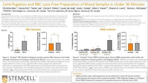 科学海报Centrifugation and RBC Lysis-Free Preparation of Blood Samples in Under 30 Minutes
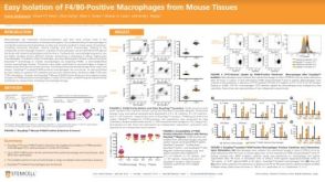 科学海报Easy Isolation of F4/80-Positive Macrophages from Mouse Tissues
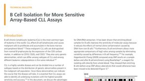 技术公告B Cell Isolation for More Sensitive Array-Based CLL Assays


 沪公网安备31010102008431号