Volpe DA and Warren MK (JUN 2003)
Toxicology in vitro : an international journal published in association with BIBRA 17 3 271--7
Myeloid clonogenic assays for comparison of the in vitro toxicity of alkylating agents.
A battery of clonal assays for myeloid progenitor cells (HPP-CFC,CFU-gemm,CFU-gm,CFU-g) was utilized to evaluate the myelotoxicity of a series of alkylating agents representing the spectrum of clinical times to nadir. Bone marrow aspirates from normal volunteers were incubated with mechlorethamine,busulfan,melphalan,carmustine or lomustine for 1 h and then cultured in methylcellulose with 30% serum and cytokines. There was a concentration-dependent inhibition of colony formation and often a differential toxicity to the myeloid progenitors with the alkylators tested. On a molar basis,mechlorethamine and melphalan were the most toxic of the alkylator drugs to the myeloid precursors. The most sensitive progenitor was CFU-gemm with the lowest inhibitory concentration IC(70) concentrations for mechlorethamine,melphalan,carmustine and lomustine. Generally,there was great similarity for drug effects between CFU-g and CFU-gm with overlapping inhibition curves. HPP-CFC proved to be the least sensitive of the progenitors to the toxic actions of the drugs. While there was no correlation between the time to clinical neutropenic nadir and the most sensitive progenitor in the clonal assays,the CFU-gm assay remains a suitable method for determining the myelotoxic potential of cytotoxic agents.
View Publication
Schiedlmeier B et al. (MAR 2003)
Blood 101 5 1759--68
High-level ectopic HOXB4 expression confers a profound in vivo competitive growth advantage on human cord blood CD34+ cells, but impairs lymphomyeloid differentiation.
Ectopic retroviral expression of homeobox B4 (HOXB4) causes an accelerated and enhanced regeneration of murine hematopoietic stem cells (HSCs) and is not known to compromise any program of lineage differentiation. However,HOXB4 expression levels for expansion of human stem cells have still to be established. To test the proposed hypothesis that HOXB4 could become a prime tool for in vivo expansion of genetically modified human HSCs,we retrovirally overexpressed HOXB4 in purified cord blood (CB) CD34+ cells together with green fluorescent protein (GFP) as a reporter protein,and evaluated the impact of ectopic HOXB4 expression on proliferation and differentiation in vitro and in vivo. When injected separately into nonobese diabetic-severe combined immunodeficient (NOD/SCID) mice or in competition with control vector-transduced cells,HOXB4-overexpressing cord blood CD34+ cells had a selective growth advantage in vivo,which resulted in a marked enhancement of the primitive CD34+ subpopulation (P =.01). However,high HOXB4 expression substantially impaired the myeloerythroid differentiation program,and this was reflected in a severe reduction of erythroid and myeloid progenitors in vitro (P textless.03) and in vivo (P =.01). Furthermore,HOXB4 overexpression also significantly reduced B-cell output (P textless.01). These results show for the first time unwanted side effects of ectopic HOXB4 expression and therefore underscore the need to carefully determine the therapeutic window of HOXB4 expression levels before initializing clinical trials.
View Publication
产品类型:
产品号#:
04434
04444
09600
09650
产品名:
MethoCult™H4434经典
MethoCult™H4434经典
StemSpan™ SFEM
StemSpan™ SFEM
Aflaki E et al. (JUN 2014)
Science translational medicine 6 240 240ra73
Macrophage models of Gaucher disease for evaluating disease pathogenesis and candidate drugs.
Gaucher disease is caused by an inherited deficiency of glucocerebrosidase that manifests with storage of glycolipids in lysosomes,particularly in macrophages. Available cell lines modeling Gaucher disease do not demonstrate lysosomal storage of glycolipids; therefore,we set out to develop two macrophage models of Gaucher disease that exhibit appropriate substrate accumulation. We used these cellular models both to investigate altered macrophage biology in Gaucher disease and to evaluate candidate drugs for its treatment. We generated and characterized monocyte-derived macrophages from 20 patients carrying different Gaucher disease mutations. In addition,we created induced pluripotent stem cell (iPSC)-derived macrophages from five fibroblast lines taken from patients with type 1 or type 2 Gaucher disease. Macrophages derived from patient monocytes or iPSCs showed reduced glucocerebrosidase activity and increased storage of glucocerebroside and glucosylsphingosine in lysosomes. These macrophages showed efficient phagocytosis of bacteria but reduced production of intracellular reactive oxygen species and impaired chemotaxis. The disease phenotype was reversed with a noninhibitory small-molecule chaperone drug that enhanced glucocerebrosidase activity in the macrophages,reduced glycolipid storage,and normalized chemotaxis and production of reactive oxygen species. Macrophages differentiated from patient monocytes or patient-derived iPSCs provide cellular models that can be used to investigate disease pathogenesis and facilitate drug development.
View Publication
产品类型:
产品号#:
19059
19059RF
85850
85857
产品名:
EasySep™人单核细胞富集试剂盒
RoboSep™ 人单核细胞富集试剂盒含滤芯吸头
mTeSR™1
mTeSR™1
Katori S et al. (JUL 2009)
The Journal of neuroscience : the official journal of the Society for Neuroscience 29 29 9137--47
Protocadherin-alpha family is required for serotonergic projections to appropriately innervate target brain areas.
Serotonergic axons from the raphe nuclei in the brainstem project to every region of the brain,where they make connections through their extensive terminal arborizations. This serotonergic innervation contributes to various normal behaviors and psychiatric disorders. The protocadherin-alpha (Pcdha) family of clustered protocadherins consists of 14 cadherin-related molecules generated from a single gene cluster. We found that the Pcdhas were strongly expressed in the serotonergic neurons. To elucidate their roles,we examined serotonergic fibers in a mouse mutant (Pcdha(Delta CR/Delta CR)) lacking the Pcdha cytoplasmic region-encoding exons,which are common to the gene cluster. In the first week after birth,the distribution pattern of serotonergic fibers in Pcdha(Delta CR/Delta CR) mice was similar to wild-type,but by 3 weeks of age,when the serotonergic axonal termini complete their arborizations,the distribution of the projections was abnormal. In some target regions,notably the globus pallidus and substantia nigra,the normally even distribution of serotonin axonal terminals was,in the mutants,dense at the periphery of each region,but sparse in the center. In the stratum lacunosum-molecular of the hippocampus,the mutants showed denser serotonergic innervation than in wild-type,and in the dentate gyrus of the hippocampus and the caudate-putamen,the innervation was sparser. Together,the abnormalities suggested that Pcdha proteins are important in the late-stage maturation of serotonergic projections. Further examination of alternatively spliced exons encoding the cytoplasmic tail showed that the A-type (but not the B-type) cytoplasmic tail was essential for the normal development of serotonergic projections.
View Publication

 EasySep™小鼠TIL(CD45)正选试剂盒


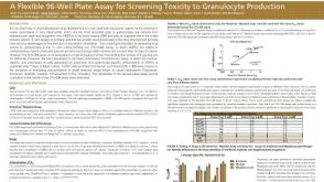 科学海报A Flexible 96-Well Plate Assay for Screening Toxicity to Granulocyte Production
 科学海报Animal Component- and Extracellular Vesicle-Free Medium Facilitates Isolation and Characterization of Extracellular Vesicles from Human Bone Marrow Mesenchymal Stromal Cells
 实验方案How to Process Leukocyte Reduction System (LRS) Cones/Chambers for Downstream Cell Isolation
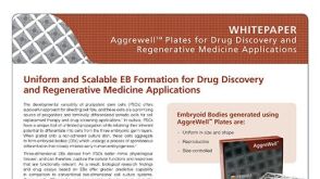 技术公告Uniform and Scalable EB Formation for Drug Discovery and Regenerative Medicine Applications


 沪公网安备31010102008431号