Ketola K et al. (DEC 2010)
Molecular cancer therapeutics 9 12 3175--85
Monensin is a potent inducer of oxidative stress and inhibitor of androgen signaling leading to apoptosis in prostate cancer cells.
Current treatment options for advanced and hormone refractory prostate cancer are limited and responses to commonly used androgen pathway inhibitors are often unsatisfactory. Our recent results indicated that sodium ionophore monensin is one of the most potent and cancer-specific inhibitors in a systematic sensitivity testing of most known drugs and drug-like molecules in a panel of prostate cancer cell models. Because monensin has been extensively used in veterinary applications to build muscle mass in cattle,the link to prostate cancer and androgen signaling was particularly interesting. Here,we showed that monensin effects at nanomolar concentrations are linked to induction of apoptosis and potent reduction of androgen receptor mRNA and protein in prostate cancer cells. Monensin also elevated intracellular oxidative stress in prostate cancer cells as evidenced by increased generation of intracellular reactive oxygen species and by induction of a transcriptional profile characteristic of an oxidative stress response. Importantly,the antiproliferative effects of monensin were potentiated by combinatorial treatment with the antiandrogens and antagonized by antioxidant vitamin C. Taken together,our results suggest monensin as a potential well-tolerated,in vivo compatible drug with strong proapoptotic effects in prostate cancer cells,and synergistic effects with antiandrogens. Moreover,our data suggest a general strategy by which the effects of antiandrogens could be enhanced by combinatorial administration with agents that increase oxidative stress in prostate cancer cells.
View Publication
Fenouille N et al. (DEC 2010)
Cancer research 70 23 9659--70
Persistent activation of the Fyn/ERK kinase signaling axis mediates imatinib resistance in chronic myelogenous leukemia cells through upregulation of intracellular SPARC.
SPARC is an extracellular matrix protein that exerts pleiotropic effects on extracellular matrix organization,growth factor availability,cell adhesion,differentiation,and immunity in cancer. Chronic myelogenous leukemia (CML) cells resistant to the BCR-ABL inhibitor imatinib (IM-R cells) were found to overexpress SPARC mRNA. In this study,we show that imatinib triggers SPARC accumulation in a variety of tyrosine kinase inhibitor (TKI)-resistant CML cell lines. SPARC silencing in IM-R cells restored imatinib sensitivity,whereas enforced SPARC expression in imatinib-sensitive cells promoted viability as well as protection against imatinib-mediated apoptosis. Notably,we found that the protective effect of SPARC required intracellular retention inside cells. Accordingly,SPARC was not secreted into the culture medium of IM-R cells. Increased SPARC expression was intimately linked to persistent activation of the Fyn/ERK kinase signaling axis. Pharmacologic inhibition of this pathway or siRNA-mediated knockdown of Fyn kinase resensitized IM-R cells to imatinib. In support of our findings,increased levels of SPARC mRNA were documented in blood cells from CML patients after 1 year of imatinib therapy compared with initial diagnosis. Taken together,our results highlight an important role for the Fyn/ERK signaling pathway in imatinib-resistant cells that is driven by accumulation of intracellular SPARC.
View Publication
产品类型:
产品号#:
04100
产品名:
MethoCult™ H4100
Aichberger KJ et al. (DEC 2009)
Blood 114 26 5342--51
Identification of proapoptotic Bim as a tumor suppressor in neoplastic mast cells: role of KIT D816V and effects of various targeted drugs.
Systemic mastocytosis (SM) is a myeloid neoplasm involving mast cells (MCs) and their progenitors. In most cases,neoplastic cells display the D816V-mutated variant of KIT. KIT D816V exhibits constitutive tyrosine kinase (TK) activity and has been implicated in increased survival and growth of neoplastic MCs. Recent data suggest that the proapoptotic BH3-only death regulator Bim plays a role as a tumor suppressor in various myeloid neoplasms. We found that KIT D816V suppresses expression of Bim in Ba/F3 cells. The KIT D816-induced down-regulation of Bim was rescued by the KIT-targeting drug PKC412/midostaurin. Both PKC412 and the proteasome-inhibitor bortezomib were found to decrease growth and promote expression of Bim in MC leukemia cell lines HMC-1.1 (D816V negative) and HMC-1.2 (D816V positive). Both drugs were also found to counteract growth of primary neoplastic MCs. Furthermore,midostaurin was found to cooperate with bortezomib and with the BH3-mimetic obatoclax in producing growth inhibition in both HMC-1 subclones. Finally,a Bim-specific siRNA was found to rescue HMC-1 cells from PKC412-induced cell death. Our data show that KIT D816V suppresses expression of proapoptotic Bim in neoplastic MCs. Targeting of Bcl-2 family members by drugs promoting Bim (re)-expression,or by BH3-mimetics such as obatoclax,may be an attractive therapy concept in SM.
View Publication
产品类型:
产品号#:
09600
09650
产品名:
StemSpan™ SFEM
StemSpan™ SFEM
Schiedlmeier B et al. (MAR 2003)
Blood 101 5 1759--68
High-level ectopic HOXB4 expression confers a profound in vivo competitive growth advantage on human cord blood CD34+ cells, but impairs lymphomyeloid differentiation.
Ectopic retroviral expression of homeobox B4 (HOXB4) causes an accelerated and enhanced regeneration of murine hematopoietic stem cells (HSCs) and is not known to compromise any program of lineage differentiation. However,HOXB4 expression levels for expansion of human stem cells have still to be established. To test the proposed hypothesis that HOXB4 could become a prime tool for in vivo expansion of genetically modified human HSCs,we retrovirally overexpressed HOXB4 in purified cord blood (CB) CD34+ cells together with green fluorescent protein (GFP) as a reporter protein,and evaluated the impact of ectopic HOXB4 expression on proliferation and differentiation in vitro and in vivo. When injected separately into nonobese diabetic-severe combined immunodeficient (NOD/SCID) mice or in competition with control vector-transduced cells,HOXB4-overexpressing cord blood CD34+ cells had a selective growth advantage in vivo,which resulted in a marked enhancement of the primitive CD34+ subpopulation (P =.01). However,high HOXB4 expression substantially impaired the myeloerythroid differentiation program,and this was reflected in a severe reduction of erythroid and myeloid progenitors in vitro (P textless.03) and in vivo (P =.01). Furthermore,HOXB4 overexpression also significantly reduced B-cell output (P textless.01). These results show for the first time unwanted side effects of ectopic HOXB4 expression and therefore underscore the need to carefully determine the therapeutic window of HOXB4 expression levels before initializing clinical trials.
View Publication

 EasySep™小鼠TIL(CD45)正选试剂盒


 科学海报Animal Component- and Extracellular Vesicle-Free Medium Facilitates Isolation and Characterization of Extracellular Vesicles from Human Bone Marrow Mesenchymal Stromal Cells
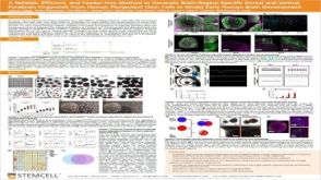 科学海报A Reliable, Efficient, and Feeder-Free Method to Generate Brain-Region-Specific Dorsal and Ventral Forebrain Organoids From Human Pluripotent Stem Cells to Model Early Human Brain Development
 沪公网安备31010102008431号