Generation of mouse monoclonal antibodies specific to Chikungunya virus using ClonaCell-HY hybridoma cloning kit
Monoclonal antibodies offer high specificity and this makes it an important tool for molecular biology,biochemistry and medicine. Typically,monoclonal antibodies are generated by fusing mouse spleen cells that have been immunized with the desired antigen with myeloma cells to create immortalized hybridomas. Here,we describe the generation of monoclonal antibodies that are specific to Chikungunya virus using ClonaCell-HY system.
View Publication
Lee WT et al. (DEC 2016)
Developmental & Comparative Immunology 65 114--123
Identification of secreted and membrane-bound bat immunoglobulin using a Microchiropteran-specific mouse monoclonal antibody
Bat immunity has received increasing attention because some bat species are being decimated by the fungal disease,White Nose Syndrome,while other species are potential reservoirs of zoonotic viruses. Identifying specific immune processes requires new specific tools and reagents. In this study,we describe a new mouse monoclonal antibody (mAb) reactive with Eptesicus fuscus immunoglobulins. The epitope recognized by mAb BT1-4F10 was localized to immunoglobulin light (lambda) chains; hence,the mAb recognized serum immunoglobulins and B lymphocytes. The BT1-4F10 epitope appeared to be restricted to Microchiropteran immunoglobulins and absent from Megachiropteran immunoglobulins. Analyses of sera and other E. fuscus fluids showed that most,if not all,secreted immunoglobulins utilized lambda light chains. Finally,mAb BT1-4F10 permitted the identification of B cell follicles in splenic white pulp. This Microchiropteran-specific mAb has potential utility in seroassays; hence,this reagent may have both basic and practical applications for studying immune process.
View Publication
产品类型:
产品号#:
03800
03801
03802
03803
03804
03805
03806
产品名:
ClonaCell™-HY 杂交瘤试剂盒
ClonaCell™-HY Medium
ClonaCell™-HY Medium
ClonaCell™-HY Medium
ClonaCell™-HY Medium
ClonaCell™-HY Medium
ClonaCell™-HY PEG (融合)
Yano M and Pirofski L-a (JAN 2011)
Clinical and vaccine immunology : CVI 18 1 59--66
Characterization of gene use and efficacy of mouse monoclonal antibodies to Streptococcus pneumoniae serotype 8.
Streptococcus pneumoniae is the most common cause of community-acquired pneumonia in the United States and globally. Despite the availability of pneumococcal capsular polysaccharide (PPS) and protein conjugate-based vaccines,the prevalence of antibiotic-resistant pneumococcal strains,serotype (ST) replacement in nonconjugate vaccine strains,and uncertainty as to whether the PPS vaccine that is used in adults protects against pneumonia emphasize the need for continued efforts to understand the nature of protective PPS antibody responses. In this study,we generated mouse monoclonal antibodies (MAbs) to a conjugate consisting of the PPS of serotype 8 (PPS8) S. pneumoniae and tetanus toxoid. Thirteen MAbs,including four IgMs that bound to PPS8 and phosphorylcholine (PC) and five IgMs and four IgG1s that bound to PPS8 but not PC,were produced,and their nucleotide sequences,epitope and fine specificity,and efficacy against lethal challenge with ST8 S. pneumoniae were determined. MAbs that bound to PPS8 exhibited gene use that was distinct from that exhibited by MAbs that bound to PC. Only PPS8-binding MAbs that did not bind PC were protective in mice. All 13 MAbs used germ line variable-region heavy (V(H)) and light (V(L)) chain genes,with no evidence of somatic hypermutation. Our data reveal a relationship between PPS specificity and V(H) gene use and MAb efficacy in mice. These findings provide insight into the relationship between antibody molecular structure and function and hold promise for the development of novel surrogates for pneumococcal vaccine efficacy.
View Publication
Amelioration of murine beta-thalassemia through drug selection of hematopoietic stem cells transduced with a lentiviral vector encoding both gamma-globin and the MGMT drug-resistance gene.
Correction of murine models of beta-thalassemia has been achieved through high-level globin lentiviral vector gene transfer into mouse hematopoietic stem cells (HSCs). However,transduction of human HSCs is less robust and may be inadequate to achieve therapeutic levels of genetically modified erythroid cells. We therefore developed a double gene lentiviral vector encoding both human gamma-globin under the transcriptional control of erythroid regulatory elements and methylguanine methyltransferase (MGMT),driven by a constitutive cellular promoter. MGMT expression provides cellular resistance to alkylator drugs,which can be administered to kill residual untransduced,diseased HSCs,whereas transduced cells are protected. Mice transplanted with beta-thalassemic HSCs transduced with a gamma-globin/MGMT vector initially had subtherapeutic levels of red cells expressing gamma-globin. To enrich gamma-globin-expressing cells,transplanted mice were treated with the alkylator agent 1,3-bis-chloroethyl-1-nitrosourea. This resulted in significant increases in the number of gamma-globin-expressing red cells and the amount of fetal hemoglobin,leading to resolution of anemia. Selection of transduced HSCs was also obtained when cells were drug-treated before transplantation. Mice that received these cells demonstrated reconstitution with therapeutic levels of gamma-globin-expressing cells. These data suggest that MGMT-based drug selection holds promise as a modality to improve gene therapy for beta-thalassemia.
View Publication
产品类型:
产品号#:
09600
09650
产品名:
StemSpan™ SFEM
StemSpan™ SFEM
Dorrell C et al. (JUN 2011)
Molecular and Cellular Endocrinology 339 1-2 144--150
Isolation of mouse pancreatic alpha, beta, duct and acinar populations with cell surface markers
Tools permitting the isolation of live pancreatic cell subsets for culture and/or molecular analysis are limited. To address this,we developed a collection of monoclonal antibodies with selective surface labeling of endocrine and exocrine pancreatic cell types. Cell type labeling specificity and cell surface reactivity were validated on mouse pancreatic sections and by gene expression analysis of cells isolated using FACS. Five antibodies which marked populations of particular interest were used to isolate and study viable populations of purified pancreatic ducts,acinar cells,and subsets of acinar cells from whole pancreatic tissue or of alpha or beta cells from isolated mouse islets. Gene expression analysis showed the presence of known endocrine markers in alpha and beta cell populations and revealed that TTR and DPPIV are primarily expressed in alpha cells whereas DGKB and GPM6A have a beta cell specific expression profile.
View Publication
产品类型:
产品号#:
03800
03801
03802
03803
03804
03805
03806
03831
产品名:
ClonaCell™-HY 杂交瘤试剂盒
ClonaCell™-HY Medium
ClonaCell™-HY Medium
ClonaCell™-HY Medium
ClonaCell™-HY Medium
ClonaCell™-HY Medium
ClonaCell™-HY PEG (融合)
ClonaCell™-HY液体帽子选择培养基
Xu H et al. (JUL 2016)
Organic & biomolecular chemistry 14 26 6179--83
Cellular thermal shift and clickable chemical probe assays for the determination of drug-target engagement in live cells.
Proof of drug-target engagement in physiologically-relevant contexts is a key pillar of successful therapeutic target validation. We developed two orthogonal technologies,the cellular thermal shift assay (CETSA) and a covalent chemical probe reporter approach (harnessing sulfonyl fluoride tyrosine labeling and subsequent click chemistry) to measure the occupancy of the mRNA-decapping scavenger enzyme DcpS by a small molecule inhibitor in live cells. Enzyme affinity determined using isothermal dose response fingerprinting (ITDRFCETSA) and the concentration required to occupy 50% of the enzyme (OC50) using the chemical probe reporter assay were very similar. In this case,the chemical probe method worked well due to the long offset kinetics of the reversible inhibitor (determined using a fluorescent dye-tagged probe). This work suggests that CETSA could become the first choice assay to determine in-cell target engagement due to its simplicity.
View Publication
产品类型:
产品号#:
70025
70025.1
70025.2
70025.3
产品名:
冻存的人外周血单个核细胞
冻存的人外周血单个核细胞
冻存的人外周血单个核细胞
冻存的人外周血单个核细胞
Murphy SV et al. (JAN 2013)
Journal of biomedical materials research. Part A 101 1 272--84
Evaluation of hydrogels for bio-printing applications.
In the United States alone,there are approximately 500,000 burn injuries that require medical treatment every year. Limitations of current treatments necessitate the development of new methods that can be applied quicker,result in faster wound regeneration,and yield skin that is cosmetically similar to undamaged skin. The development of new hydrogel biomaterials and bioprinting deposition technologies has provided a platform to address this need. Herein we evaluated characteristics of twelve hydrogels to determine their suitability for bioprinting applications. We chose hydrogels that are either commercially available,or are commonly used for research purposes. We evaluated specific hydrogel properties relevant to bioprinting applications,specifically; gelation time,swelling or contraction,stability,biocompatibility and printability. Further,we described regulatory,commercial and financial aspects of each of the hydrogels. While many of the hydrogels screened may exhibit characteristics suitable for other applications,UV-crosslinked Extracel,a hyaluronic acid-based hydrogel,had many of the desired properties for our bioprinting application. Taken together with commercial availability,shelf life,potential for regulatory approval and ease of use,these materials hold the potential to be further developed into fast and effective wound healing treatments.
View Publication

 EasySep™小鼠TIL(CD45)正选试剂盒


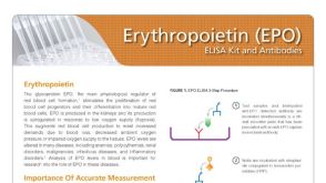 产品手册High Affinity Anti-EPO Reagents for Consistent Results


 沪公网安备31010102008431号