Badr CE et al. (MAY 2013)
JNCI: Journal of the National Cancer Institute 105 9 643--653
Targeting Cancer Cells With the Natural Compound Obtusaquinone
BACKGROUND Tumor cells present high levels of oxidative stress. Cancer therapeutics exploiting such biochemical changes by increasing reactive oxygen species (ROS) production or decreasing intracellular ROS scavengers could provide a powerful treatment strategy. METHODS To test the effect of our compound,obtusaquinone (OBT),we used several cell viability assays on seven different glioblastoma (GBM) cell lines and primary cells and on 12 different cell lines representing various cancer types in culture as well as on subcutaneous (n = 7 mice per group) and two intracranial GBM (n = 6-8 mice per group) and breast cancer (n = 6 mice per group) tumor models in vivo. Immunoblotting,immunostaining,flow cytometry,and biochemical assays were used to investigate the OBT mechanism of action. Histopathological analysis (n = 2 mice per group) and blood chemistry (n = 2 mice per group) were used to test for any compound-related toxicity. Statistical tests were two-sided. RESULTS OBT induced rapid increase in intracellular ROS levels,downregulation of cellular glutathione levels and increase in its oxidized form,and activation of cellular stress pathways and DNA damage,subsequently leading to apoptosis. Oxidative stress is believed to be the main mechanism through which this compounds targets cancer cells. OBT was well tolerated in mice,slowed tumor growth,and statistically prolonged survival in GBM tumor models. The ratio of median survival in U251 intracranial model in OBT vs control was 1.367 (95% confidence interval [CI] of ratio = 1.031 to 1.367,P = .008). Tumor growth inhibition was also observed in a mouse breast cancer model (average tumor volume per mouse,OBT vs control: 36.3 vs 200.4mm(3),difference = 164.1mm(3),95% CI =72.6 to 255.6mm(3),P = .005). CONCLUSIONS Given its properties and efficacy in cancer killing,our results suggest that OBT is a promising cancer therapeutic.
View Publication
产品类型:
产品号#:
05750
05751
产品名:
NeuroCult™ NS-A 基础培养基(人)
NeuroCult™ NS-A 扩增试剂盒(人)
Donangelo I et al. (JAN 2014)
Endocrine Related Cancer 21 2 203--216
Sca1+ murine pituitary adenoma cells show tumor-growth advantage
The role of tumor stem cells in benign tumors such as pituitary adenomas remains unclear. In this study,we investigated whether the cells within pituitary adenomas that spontaneously develop in Rb+/- mice are hierarchically distributed with a subset being responsible for tumor growth. Cells derived directly from such tumors grew as spheres in serum-free culture medium supplemented with epidermal growth factor and basic fibroblast growth factor. Some cells within growing pituitary tumor spheres (PTS) expressed common stem cell markers (Sca1,Sox2,Nestin,and CD133),but were devoid of hormone-positive differentiated cells. Under subsequent differentiating conditions (matrigel-coated growth surface),PTS expressed all six pituitary hormones. We next searched for specific markers of the stem cell population and isolated a Sca1(+) cell population that showed increased sphere formation potential,lower mRNA hormone expression,higher expression of stem cell markers (Notch1,Sox2,and Nestin),and increased proliferation rates. When transplanted into non-obese diabetic-severe combined immunodeficiency gamma mice brains,Sca1(+) pituitary tumor cells exhibited higher rates of tumor formation (brain tumors observed in 11/11 (100%) vs 7/12 (54%) of mice transplanted with Sca1(+) and Sca1(-) cells respectively). Magnetic resonance imaging and histological analysis of brain tumors showed that tumors derived from Sca1(+) pituitary tumor cells were also larger and plurihormonal. Our findings show that Sca1(+) cells derived from benign pituitary tumors exhibit an undifferentiated expression profile and tumor-proliferative advantages,and we propose that they could represent putative pituitary tumor stem/progenitor cells.
View Publication
Ostrakhovitch EA et al. (DEC 2012)
Archives of biochemistry and biophysics 528 1 21--31
Directed differentiation of embryonic P19 cells and neural stem cells into neural lineage on conducting PEDOT-PEG and ITO glass substrates.
Differentiation of pluripotent and lineage restricted stem cells such as neural stem cells (NSCs) was studied on conducting substrates of various nature without perturbation of the genome with exogenous genetic material or chemical stimuli. Primary mouse adult neural stem cells (NSCs) and P19 pluripotent embryonal (P19 EC) carcinoma cells were used. Expression levels of neuronal markers β-III-tubulin and neurofilament were evaluated by immunochemistry and flow cytometry. It was shown that the ability of the substrate to induce differentiation directly correlated with its conductivity. Conducting substrates (conducting oxides or doped pi-conjugated organic polymers) with different morphology,structure,and conductivity mechanisms all promoted differentiation of NSC and P19 cells into neuronal lineage to a similar degree without use of additional factors such as poly-L-ornithine coating or retinoic acid,as verified by their morphology and upregulation of the neuronal markers but not astrocyte marker GFAP. However,substrates with low conductance below ca. 10(-4) S cm(-2) did not show this ability. Morphology of differentiating cells was visualized by atomic force microscopy. NSCs cells increased β-III-tubulin expression by 95% and P19 cells by over 30%. Our results suggest that the substrate conductivity is a key factor governing the cell fate. Differentiation of P19 cells into neuronal lineage on conducting substrates was attributed to downregualtion of Akt signaling pathway and increase in expression of dual oxidase 1 (DUOX 1).
View Publication
产品类型:
产品号#:
05700
05701
05702
05703
05704
05715
产品名:
NeuroCult™ 基础培养基(小鼠&大鼠)
NeuroCult™ 扩增添加物 (小鼠&大鼠)
NeuroCult™ 扩增试剂盒 (小鼠&大鼠)
NeuroCult™ 分化添加物 (小鼠&大鼠)
NeuroCult™ 分化试剂盒 (小鼠&大鼠)
NeuroCult™成年中枢神经系统(CNS)组织酶解试剂盒(小鼠和大鼠)
Zhu TS et al. (SEP 2011)
Cancer research 71 18 6061--72
Endothelial cells create a stem cell niche in glioblastoma by providing NOTCH ligands that nurture self-renewal of cancer stem-like cells.
One important function of endothelial cells in glioblastoma multiforme (GBM) is to create a niche that helps promote self-renewal of cancer stem-like cells (CSLC). However,the underlying molecular mechanism for this endothelial function is not known. Since activation of NOTCH signaling has been found to be required for propagation of GBM CSLCs,we hypothesized that the GBM endothelium may provide the source of NOTCH ligands. Here,we report a corroboration of this concept with a demonstration that NOTCH ligands are expressed in endothelial cells adjacent to NESTIN and NOTCH receptor-positive cancer cells in primary GBMs. Coculturing human brain microvascular endothelial cells (hBMEC) or NOTCH ligand with GBM neurospheres promoted GBM cell growth and increased CSLC self-renewal. Notably,RNAi-mediated knockdown of NOTCH ligands in hBMECs abrogated their ability to induce CSLC self-renewal and GBM tumor growth,both in vitro and in vivo. Thus,our findings establish that NOTCH activation in GBM CSLCs is driven by juxtacrine signaling between tumor cells and their surrounding endothelial cells in the tumor microenvironment,suggesting that targeting both CSLCs and their niche may provide a novel strategy to deplete CSLCs and improve GBM treatment.
View Publication
产品类型:
产品号#:
05750
05751
05752
产品名:
NeuroCult™ NS-A 基础培养基(人)
NeuroCult™ NS-A 扩增试剂盒(人)
NeuroCult™ NS-A 分化试剂盒 (人)
Fè et al. ( 2014)
PloS one 9 3 e91519
Comparative expression study of the endo-G protein coupled receptor (GPCR) repertoire in human glioblastoma cancer stem-like cells, U87-MG cells and non malignant cells of neural origin unveils new potential therapeutic targets.
Glioblastomas (GBMs) are highly aggressive,invasive brain tumors with bad prognosis and unmet medical need. These tumors are heterogeneous being constituted by a variety of cells in different states of differentiation. Among these,cells endowed with stem properties,tumor initiating/propagating properties and particularly resistant to chemo- and radiotherapies are designed as the real culprits for tumor maintenance and relapse after treatment. These cells,termed cancer stem-like cells,have been designed as prominent targets for new and more efficient cancer therapies. G-protein coupled receptors (GPCRs),a family of membrane receptors,play a prominent role in cell signaling,cell communication and crosstalk with the microenvironment. Their role in cancer has been highlighted but remains largely unexplored. Here,we report a descriptive study of the differential expression of the endo-GPCR repertoire in human glioblastoma cancer stem-like cells (GSCs),U-87 MG cells,human astrocytes and fetal neural stem cells (f-NSCs). The endo-GPCR transcriptome has been studied using Taqman Low Density Arrays. Of the 356 GPCRs investigated,138 were retained for comparative studies between the different cell types. At the transcriptomic level,eight GPCRs were specifically expressed/overexpressed in GSCs. Seventeen GPCRs appeared specifically expressed in cells with stem properties (GSCs and f-NSCs). Results of GPCR expression at the protein level using mass spectrometry and proteomic analysis are also presented. The comparative GPCR expression study presented here gives clues for new pathways specifically used by GSCs and unveils novel potential therapeutic targets.
View Publication
Gallo M et al. (JAN 2013)
Cancer Research 73 1 417--427
A Tumorigenic MLL-Homeobox Network in Human Glioblastoma Stem Cells
Glioblastoma growth is driven by cancer cells that have stem cell properties,but molecular determinants of their tumorigenic behavior are poorly defined. In cancer,altered activity of the epigenetic modifiers Polycomb and Trithorax complexes may contribute to the neoplastic phenotype. Here,we provide the first mechanistic insights into the role of the Trithorax protein mixed lineage leukemia (MLL) in maintaining cancer stem cell characteristics in human glioblastoma. We found that MLL directly activates the Homeobox gene HOXA10. In turn,HOXA10 activates a downstream Homeobox network and other genes previously characterized for their role in tumorigenesis. The MLL-Homeobox axis we identified significantly contributes to the tumorigenic potential of glioblastoma stem cells. Our studies suggest a role for MLL in contributing to the epigenetic heterogeneity between tumor-initiating and non-tumor-initiating cells in glioblastoma.
View Publication
产品类型:
产品号#:
05750
产品名:
NeuroCult™ NS-A 基础培养基(人)
Kerosuo L et al. (DEC 2008)
Journal of cell science 121 Pt 23 3941--50
Myc increases self-renewal in neural progenitor cells through Miz-1.
The mechanisms underlying the decision of a stem or progenitor cell to either self-renew or differentiate are incompletely understood. To address the role of Myc in this process,we expressed different forms of the proto-oncogene Myc in multipotent neural progenitor cells (NPCs) using retroviral transduction. Expression of Myc in neurospheres increased the proportion of self-renewing cells fivefold,and 1% of the Myc-overexpressing cells,but none of the control cells,retained self-renewal capacity even under differentiation-inducing conditions. A Myc mutant (MycV394D) deficient in binding to Miz-1,did not increase the percentage of self-renewing cells but was able to stimulate proliferation of NPCs as efficiently as wild-type Myc,indicating that these two cellular phenomena are regulated by at least partially different pathways. Our results suggest that Myc,through Miz-1,enhances self-renewal of NPCs and influences the way progenitor cells react to the environmental cues that normally dictate the cellular identity of tissues containing self-renewing cells.
View Publication
产品类型:
产品号#:
05707
产品名:
NeuroCult™化学解离试剂盒(小鼠)
Pang ZP et al. (AUG 2011)
Nature 476 7359 220--3
Induction of human neuronal cells by defined transcription factors.
Somatic cell nuclear transfer,cell fusion,or expression of lineage-specific factors have been shown to induce cell-fate changes in diverse somatic cell types. We recently observed that forced expression of a combination of three transcription factors,Brn2 (also known as Pou3f2),Ascl1 and Myt1l,can efficiently convert mouse fibroblasts into functional induced neuronal (iN) cells. Here we show that the same three factors can generate functional neurons from human pluripotent stem cells as early as 6 days after transgene activation. When combined with the basic helix-loop-helix transcription factor NeuroD1,these factors could also convert fetal and postnatal human fibroblasts into iN cells showing typical neuronal morphologies and expressing multiple neuronal markers,even after downregulation of the exogenous transcription factors. Importantly,the vast majority of human iN cells were able to generate action potentials and many matured to receive synaptic contacts when co-cultured with primary mouse cortical neurons. Our data demonstrate that non-neural human somatic cells,as well as pluripotent stem cells,can be converted directly into neurons by lineage-determining transcription factors. These methods may facilitate robust generation of patient-specific human neurons for in vitro disease modelling or future applications in regenerative medicine.
View Publication
产品类型:
产品号#:
85850
85857
产品名:
mTeSR™1
mTeSR™1
Saito T et al. (JUL 2013)
PLoS ONE 8 7 e70010
Metformin, a Diabetes Drug, Eliminates Tumor-Initiating Hepatocellular Carcinoma Cells
Metformin has been widely used as an oral drug for diabetes mellitus for approximately 60 years. Interestingly,recent reports showed that metformin exhibited an anti-tumor action in a wide range of malignancies including hepatocellular carcinoma (HCC). In the present study,we investigated its impact on tumor-initiating HCC cells. Metformin suppressed cell growth and induced apoptosis in a dose-dependent manner. Flow cytometric analysis showed that metformin treatment markedly reduced the number of tumor-initiating epithelial cell adhesion molecule (EpCAM)(+) HCC cells. Non-adherent sphere formation assays of EpCAM(+) cells showed that metformin impaired not only their sphere-forming ability,but also their self-renewal capability. Consistent with this,immunostaining of spheres revealed that metformin significantly decreased the number of component cells positive for hepatic stem cell markers such as EpCAM and α-fetoprotein. In a xenograft transplantation model using non-obese diabetic/severe combined immunodeficient mice,metformin and/or sorafenib treatment suppressed the growth of tumors derived from transplanted HCC cells. Notably,the administration of metformin but not sorafenib decreased the number of EpCAM(+) cells and impaired their self-renewal capability. As reported,metformin activated AMP-activated protein kinase (AMPK) through phosphorylation; however its inhibitory effect on the mammalian target of rapamycin (mTOR) pathway did not necessarily correlate with its anti-tumor activity toward EpCAM(+) tumor-initiating HCC cells. These results indicate that metformin is a promising therapeutic agent for the elimination of tumor-initiating HCC cells and suggest as-yet-unknown functions other than its inhibitory effect on the AMPK/mTOR pathway.
View Publication

 EasySep™小鼠TIL(CD45)正选试剂盒


 科学海报Scale-up of Human Pluripotent Stem Cells and Differentiation to Megakaryocytes or Neural Crest Cells in 3D Suspension Culture
 技术公告Identification of ALDH-Expressing Cancer Stem Cells Using ALDEFLUOR™
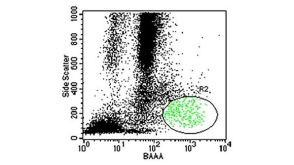

 沪公网安备31010102008431号