Senatus PB et al. (JAN 2006)
Molecular cancer therapeutics 5 1 20--8
Restoration of p53 function for selective Fas-mediated apoptosis in human and rat glioma cells in vitro and in vivo by a p53 COOH-terminal peptide.
We have shown that a COOH-terminal peptide of p53 (amino acids 361-382,p53p),linked to the truncated homeobox domain of Antennapedia (Ant) as a carrier for transduction,induced rapid apoptosis in human premalignant and malignant cell lines. Here,we report that human and rat glioma lines containing endogenous mutant p53 or wild-type (WT) p53 were induced into apoptosis by exposure to this peptide called p53p-Ant. The peptide was comparatively nontoxic to proliferating nonmalignant human and rat glial cell lines containing WT p53 and proliferating normal human peripheral marrow blood stem cells. Degree of sensitivity to the peptide correlated directly with the level of endogenous p53 expression and mutant p53 conformation. Apoptosis induction by p53p-Ant was quantitated by terminal deoxynucleotidyl transferase-mediated dUTP nick end labeling assay and Annexin V staining in human glioma cells in vitro and in a syngeneic orthotopic 9L glioma rat model using convection-enhanced delivery in vivo. The mechanism of cell death by this peptide was solely through the Fas extrinsic apoptotic pathway. p53p-Ant induced a 3-fold increase in extracellular membrane Fas expression in glioma cells but no significant increase in nonmalignant glial cells. These data suggest that p53 function for inducing Fas-mediated apoptosis in gliomas,which express sufficient quantities of endogenous mutant or WT p53,may be restored or activated,respectively,by a cell-permeable peptide derived from the p53 COOH-terminal regulatory domain (p53p-Ant). p53p-Ant may serve as a prototypic model for the development of new anticancer agents with unique selectivity for glioma cancer cells and it can be successfully delivered in vivo into a brain tumor by a convection-enhanced delivery system,which circumvents the blood-brain barrier.
View Publication
产品类型:
产品号#:
04434
04444
产品名:
MethoCult™H4434经典
MethoCult™H4434经典
Lambert AA et al. (AUG 2008)
Blood 112 4 1299--307
The C-type lectin surface receptor DCIR acts as a new attachment factor for HIV-1 in dendritic cells and contributes to trans- and cis-infection pathways.
The dynamic interplay between dendritic cells (DCs) and human immunodeficiency virus type-1 (HIV-1) is thought to result in viral dissemination and evasion of antiviral immunity. Although initial observations suggested that the C-type lectin receptor (CLR) DC-SIGN was responsible for the trans-infection function of the virus,subsequent studies demonstrated that trans-infection of CD4(+) T cells with HIV-1 can also occur through DC-SIGN-independent mechanisms. We demonstrate that a cell surface molecule designated DCIR (for DC immunoreceptor),a member of a recently described family of DC-expressing CLRs,can participate in the capture of HIV-1 and promote infection in trans and in cis of autologous CD4(+) T cells from human immature monocyte-derived DCs. The contribution of DCIR to these processes was revealed using DCIR-specific siRNAs and a polyclonal antibody specific for the carbohydrate recognition domain of DCIR. Data from transfection experiments indicated that DCIR acts as a ligand for HIV-1 and is involved in events leading to productive virus infection. Finally,we show that the neck domain of DCIR is important for the DCIR-mediated effect on virus binding and infection. These results point to a possible role for DCIR in HIV-1 pathogenesis by supporting the productive infection of DCs and promoting virus propagation.
View Publication
产品类型:
产品号#:
19052
19052RF
产品名:
EasySep™人CD4+ T细胞富集试剂盒
RoboSep™ 人CD4+ T细胞富集试剂盒含滤芯吸头
Kwok CTD et al. (MAR 2016)
Stem Cell Research 16 3 651--661
The Forkhead box transcription factor FOXM1 is required for the maintenance of cell proliferation and protection against oxidative stress in human embryonic stem cells
Human embryonic stem cells (hESCs) exhibit unique cell cycle structure,self-renewal and pluripotency. The Forkhead box transcription factor M1 (FOXM1) is critically required for the maintenance of pluripotency in mouse embryonic stem cells and mouse embryonal carcinoma cells,but its role in hESCs remains unclear. Here,we show that FOXM1 expression was enriched in undifferentiated hESCs and was regulated in a cell cycle-dependent manner with peak levels detected at the G2/M phase. Expression of FOXM1 did not correlate with OCT4 and NANOG during in vitro differentiation of hESCs. Importantly,knockdown of FOXM1 expression led to aberrant cell cycle distribution with impairment in mitotic progression but showed no profound effect on the undifferentiated state. Interestingly,FOXM1 depletion sensitized hESCs to oxidative stress. Moreover,genome-wide analysis of FOXM1 targets by ChIP-seq identified genes important for M phase including CCNB1 and CDK1,which were subsequently confirmed by ChIP and RNA interference analyses. Further peak set comparison against a differentiating hESC line and a cancer cell line revealed a substantial difference in the genomic binding profile of FOXM1 in hESCs. Taken together,our findings provide the first evidence to support FOXM1 as an important regulator of cell cycle progression and defense against oxidative stress in hESCs.
View Publication
产品类型:
产品号#:
05110
85850
85857
产品名:
STEMdiff™定型内胚层检测试剂盒
mTeSR™1
mTeSR™1
(Apr 2025)
Cells 14 8
LFA-1/ICAM-1 Interactions Between CD8+ and CD4+ T Cells Promote CD4+ Th1-Dominant Differentiation and CD8+ T Cell Cytotoxicity for Strong Antitumor Immunity After Cryo-Thermal Therapy
CD4+ T cells have been well-regarded as “helper” cells in activating the cytotoxicity of CD8+ T cells for effective tumor eradication,while few studies have focused on whether CD8+ T cells regulate CD4+ T cells. Our previous studies provided evidence for an interaction between CD4+ and CD8+ T cells after cryo-thermal therapy,but the mechanism remains unclear,especially pertaining to how CD8+ T cells promote the Th1 differentiation of CD4+ T cells. This study revealed that activated CD4+ and CD8+ T cells are critical for CTT-induced antitumor immunity,and the interaction between activated T cells is enhanced. The reciprocal regulation of activated CD8+ and CD4+ T cells was through LFA-1/ICAM-1 interactions,in which CD8+ T cells facilitate Notch1-dependent CD4+ Th1-dominant differentiation and promote IL-2 secretion of CD4+ T cells. Meanwhile,IL-2 derived from CD4+ T cells enhances the cytotoxicity of CD8+ T cells and establishes a positive feedback loop via increasing the expression of LFA-1 and ICAM-1 on T cells. Clinical analyses further validated that LFA-1/ICAM interactions between CD4+ and CD8+ T cells are correlated with clinical outcomes. Our study extends the functions of the LFA-1/ICAM-1 adhesion pathway,indicating its novel role in the interaction of CD4+ and CD8+ T cells.
View Publication
Ng Y-S et al. (OCT 2004)
The Journal of experimental medicine 200 7 927--34
Bruton's tyrosine kinase is essential for human B cell tolerance.
Most polyreactive and antinuclear antibodies are removed from the human antibody repertoire during B cell development. To elucidate how B cell receptor (BCR) signaling may regulate human B cell tolerance,we tested the specificity of recombinant antibodies from single peripheral B cells isolated from patients suffering from X-linked agammaglobulinemia (XLA). These patients carry mutations in the Bruton's tyrosine kinase (BTK) gene that encode an essential BCR signaling component. We find that in the absence of Btk,peripheral B cells show a distinct antibody repertoire consistent with extensive secondary V(D)J recombination. Nevertheless,XLA B cells are enriched in autoreactive clones. Our results demonstrate that Btk is essential in regulating thresholds for human B cell tolerance.
View Publication
产品类型:
产品号#:
15024
15064
产品名:
RosetteSep™ 人B细胞富集抗体混合物
RosetteSep™人B细胞富集抗体混合物
Elliott DA et al. (DEC 2011)
Nature methods 8 12 1037--1040
NKX2-5(eGFP/w) hESCs for isolation of human cardiac progenitors and cardiomyocytes.
NKX2-5 is expressed in the heart throughout life. We targeted eGFP sequences to the NKX2-5 locus of human embryonic stem cells (hESCs); NKX2-5(eGFP/w) hESCs facilitate quantification of cardiac differentiation,purification of hESC-derived committed cardiac progenitor cells (hESC-CPCs) and cardiomyocytes (hESC-CMs) and the standardization of differentiation protocols. We used NKX2-5 eGFP(+) cells to identify VCAM1 and SIRPA as cell-surface markers expressed in cardiac lineages.
View Publication
OrganoLabeler: A Quick and Accurate Annotation Tool for Organoid Images
Organoids are self-assembled 3D cellular structures that resemble organs structurally and functionally,providing in vitro platforms for molecular and therapeutic studies. Generation of organoids from human cells often requires long and costly procedures with arguably low efficiency. Prediction and selection of cellular aggregates that result in healthy and functional organoids can be achieved by using artificial intelligence-based tools. Transforming images of 3D cellular constructs into digitally processable data sets for training deep learning models requires labeling of morphological boundaries,which often is performed manually. Here,we report an application named OrganoLabeler,which can create large image-based data sets in a consistent,reliable,fast,and user-friendly manner. OrganoLabeler can create segmented versions of images with combinations of contrast adjusting,K-means clustering,CLAHE,binary,and Otsu thresholding methods. We created embryoid body and brain organoid data sets,of which segmented images were manually created by human researchers and compared with OrganoLabeler. Validation is performed by training U-Net models,which are deep learning models specialized in image segmentation. U-Net models,which are trained with images segmented by OrganoLabeler,achieved similar or better segmentation accuracies than the ones trained with manually labeled reference images. OrganoLabeler can replace manual labeling,providing faster and more accurate results for organoid research free of charge.
View Publication
产品类型:
产品号#:
100-0483
100-0484
85850
85857
产品名:
Hausser Scientificᵀᴹ 明线血球计数板
ReLeSR™
mTeSR™1
mTeSR™1
M. K. Orlowska et al. (Mar 2024)
Biomicrofluidics 18 2
A miniaturized culture platform for control of the metabolic environment
The heart is a metabolic “omnivore” and adjusts its energy source depending on the circulating metabolites. Human cardiac organoids,a three-dimensional in vitro model of the heart wall,are a useful tool to study cardiac physiology and pathology. However,cardiac tissue naturally experiences shear stress and nutrient fluctuations via blood flow in vivo,whilst in vitro models are conventionally cultivated in a static medium. This necessitates the regular refreshing of culture media,which creates acute cellular disturbances and large metabolic fluxes. To culture human cardiac organoids in a more physiological manner,we have developed a perfused bioreactor for cultures in a 96-well plate format. The designed bioreactor is easy to fabricate using a common culture plate and a 3D printer. Its open system allows for the use of traditional molecular biology techniques,prevents flow blockage issues,and provides easy access for sampling and cell assays. We hypothesized that a perfused culture would create more stable environment improving cardiac function and maturation. We found that lactate is rapidly produced by human cardiac organoids,resulting in large fluctuations in this metabolite under static culture. Despite this,neither medium perfusion in bioreactor culture nor lactate supplementation improved cardiac function or maturation. In fact,RNA sequencing revealed little change across the transcriptome. This demonstrates that cardiac organoids are robust in response to fluctuating environmental conditions under normal physiological conditions. Together,we provide a framework for establishing an easily accessible perfusion system that can be adapted to a range of miniaturized cell culture systems.
View Publication

 EasySep™小鼠TIL(CD45)正选试剂盒


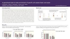 科学海报Fast and Easy Enrichment of Cell Subsets for HLA Analysis
 30:53
线上讲座Highly Characterized Human iPSCs and NPCs for Downstream Differentiation Applications发布日期: 07/19/2023
30:53
线上讲座Highly Characterized Human iPSCs and NPCs for Downstream Differentiation Applications发布日期: 07/19/2023

 沪公网安备31010102008431号