
 EasySep™小鼠TIL(CD45)正选试剂盒


搜索结果: 'methocult media formulations for mouse hematopoietic cells serum containing'
-

产品类型:
产品号#:
19852
19852RF
18783
18783RF
18765
18765RF
产品名:
EasySep™小鼠CD4+ T细胞分选试剂盒
RoboSep™ 小鼠CD4+ T细胞分选试剂盒
EasySep™小鼠CD4+CD25+调节性T细胞分选试剂盒II
RoboSep™ 小鼠CD4+CD25+调节性T细胞分选试剂盒II
EasySep™小鼠CD4+ CD62L+ T细胞分选试剂盒
RoboSep™ 小鼠CD4+ CD62L+ T细胞分选试剂盒
-
产品类型:
产品号#:
09600
09650
产品名:
StemSpan™ SFEM
StemSpan™ SFEM
-
产品类型:
产品号#:
100-0276
100-1130
产品名:
mTeSR™ Plus
mTeSR™ Plus
-
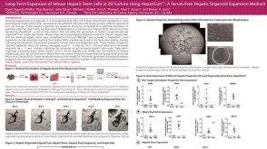 科学海报Long-Term Expansion of Mouse Hepatic Stem Cells in 3D Culture Using HepatiCult™: A Serum-Free Hepatic Organoid Expansion Medium
产品类型:
Conference:
ISSCR 2017
产品号#:
70932
产品名:
-
产品类型:
产品号#:
05850
05857
05870
05875
05872
05873
07909
07920
85850
85857
85870
85875
100-0483
100-0484
07922
产品名:
IV型胶原酶(1mg /mL)
ACCUTASE™
mTeSR™1
mTeSR™1
Hausser Scientificᵀᴹ 明线血球计数板
ReLeSR™
ACCUTASE™
-
产品类型:
产品号#:
05850
05857
05870
05875
85850
85857
85870
85875
产品名:
mTeSR™1
mTeSR™1
-
产品类型:
产品号#:
04436
04064
04100
04230
04236
04431
04434
04444
04464
04531
04535
04545
04536
04564
04035
04330
04034
04044
04435
04445
04534
04544
04437
04447
产品名:
MethoCult™ SF H4436
MethoCult™ H4034 Optimum 入门试剂盒
MethoCult™ H4100
MethoCult™ H4230
MethoCult™ SF H4236
MethoCult™ H4431
MethoCult™ H4434 Classic
MethoCult™ H4434 Classic
MethoCult™ H4434 Classic 套装
MethoCult™ H4531
MethoCult™ H4535 Enriched,不含EPO
MethoCult™ H4535 Enriched,不含EPO
MethoCult™ SF H4536
MethoCult™ H4534 Classic 无 EPO 入门试剂盒
MethoCult™ 不含EPO的H4035 Optimum
MethoCult™ H4330
MethoCult™ H4034 Optimum
MethoCult™ H4034 Optimum
MethoCult™ H4435 Enriched
MethoCult™ H4435 Enriched
MethoCult™ H4534 Classic(不含 EPO)
MethoCult™ H4534 Classic(不含 EPO)
MethoCult™ Express
MethoCult™ Express
-
产品类型:
产品号#:
03434
03444
03236
产品名:
MethoCult™ GF M3434
MethoCult™ GF M3434
MethoCult™ SF M3236
-
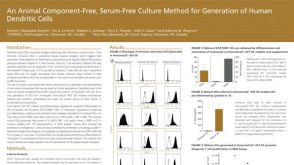 科学海报An Animal Component-Free, Serum-Free Culture Method for Generation of Human Dendritic Cells
产品类型:
Conference:
AAI 2016
产品号#:
10985
10986
10987
10988
10989
产品名:
ImmunoCult™ 树突状细胞培养试剂盒
ImmunoCult™-ACF树突状细胞培养基
ImmunoCult™-ACF树突状细胞培养基
ImmunoCult™-ACF树突状细胞分化添加物
ImmunoCult™树突状细胞成熟添加物
发布日期: 07/20/2016 -
产品类型:
产品号#:
04435
04445
05150
产品名:
MethoCult™ H4435 Enriched
MethoCult™ H4435 Enriched
MyeloCult™ H5100
-

 沪公网安备31010102008431号