
 EasySep™小鼠TIL(CD45)正选试剂盒


产品号 #03814_C
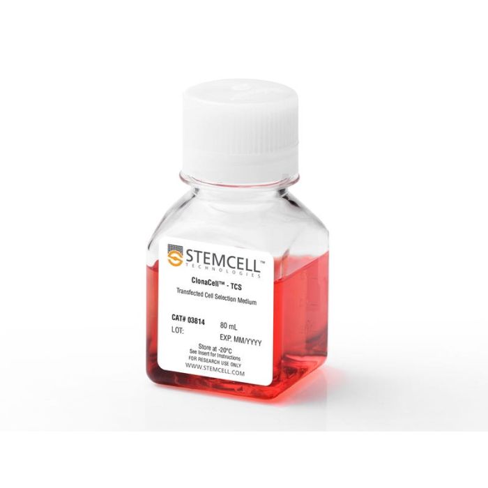
用于筛选和克隆悬浮适应性细胞和贴壁细胞的半固体甲基纤维素基培养基(含血清)
若您需要咨询产品或有任何技术问题,请通过官方电话 400 885 9050 或邮箱 info.cn@stemcell.com 与我们联系。
用于筛选和克隆悬浮适应性细胞和贴壁细胞的半固体甲基纤维素基培养基(含血清)
ClonaCell™-TCS 培养基是一款含血清、基于甲基纤维素的半固体培养基,适用于筛选和克隆多种悬浮适应性细胞株,包括 CHO-S 和杂交瘤细胞。该培养基亦可用于部分贴壁生长、在血清条件下培养的细胞系的半固体克隆培养,如 CHO-K1、BHK-1 和 HEK-293 等。该培养基含经过预筛选的胎牛血清(FBS)和牛血清白蛋白(BSA),可支持多种细胞类型的稳健生长。ClonaCell™-TCS 培养基不含筛选剂。
半固体克隆培养的优势:
•单个细胞悬浮于高黏度培养基中,形成物理分离、独立的克隆集落,便于挑取;
•相较于极限稀释法,使用该方法可更快速、节省资源地获得单克隆细胞系;
•在黏性培养基中,具有不同生长速率和产量的多样性克隆均可形成独立集落,从而更易于筛选出稀有的高产克隆。相比液体批量培养,半固体培养支持同时进行筛选和克隆,提高高表达克隆的获取效率。
包含
• IMDM (Iscove's改良杜氏培养基)
• 甲基纤维素
• 预筛选血清
• 牛血清白蛋白
• 2-巯基乙醇
• 酚红
• L-谷氨酰胺
• 其他成分
分类
半固体培养基,专用培养基
细胞类型
CHO细胞,HEK-293细胞(人胚肾293细胞),杂交瘤细胞,其他物种
种属
小鼠,其他物种
应用
细胞培养,半固体克隆
品牌
ClonaCell
研究领域
抗体制备,细胞系制备,杂交瘤制备
请在《产品说明书》中查找相关支持信息和使用说明,或浏览下方更多实验方案。
本产品专为以下研究领域设计,适用于工作流程中的高亮阶段。探索这些工作流程,了解更多我们为各研究领域提供的其他配套产品。
| 物种 | 其它物种, 小鼠 |
|---|---|
| Contains | • IMDM (Iscove's Modified Dulbecco's Medium) • Methylcellulose • Pre-selected serum • Bovine serum albumin • 2-Mercaptoethanol • Phenol red • L-Glutamine • Other ingredients |

用于筛选转染的哺乳动物细胞的抗生素
用于筛选转染后原核和真核细胞的抗生素
在线联系


 沪公网安备31010102008431号