
 EasySep™小鼠TIL(CD45)正选试剂盒


产品号 #100-0500_C
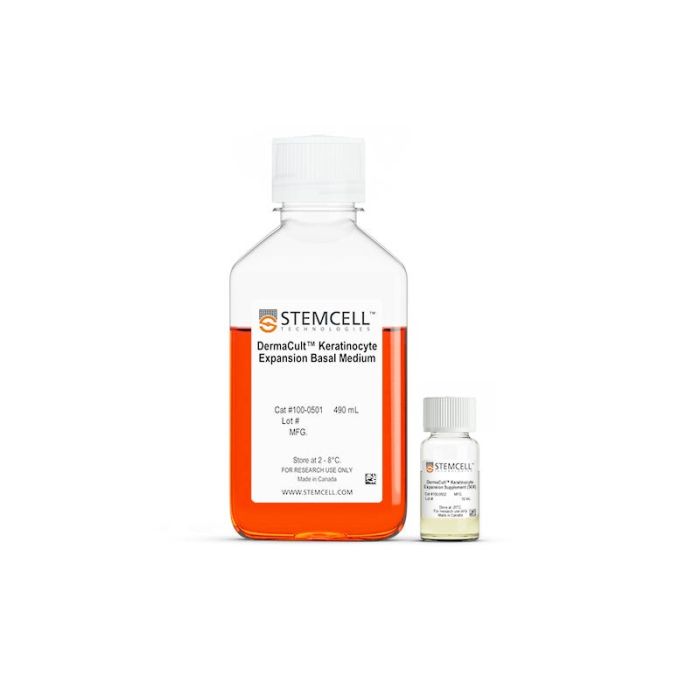
人原代角质形成细胞的无血清和无BPE扩增培养基
若您需要咨询产品或有任何技术问题,请通过官方电话 400 885 9050 或邮箱 info.cn@stemcell.com 与我们联系。
人原代角质形成细胞的无血清和无BPE扩增培养基
使用无血清、无牛垂体提取物(BPE)的 DermaCult™ 角质形成细胞扩增培养基,无需饲养层,可显著提升人原代角质形成细胞的衍生效率和扩增速率,实现长期培养。细胞扩增更高效,意味着您可以从同一样本中进行多次重复实验及技术重复,无需反复购买未分化细胞或重新获取患者样本。
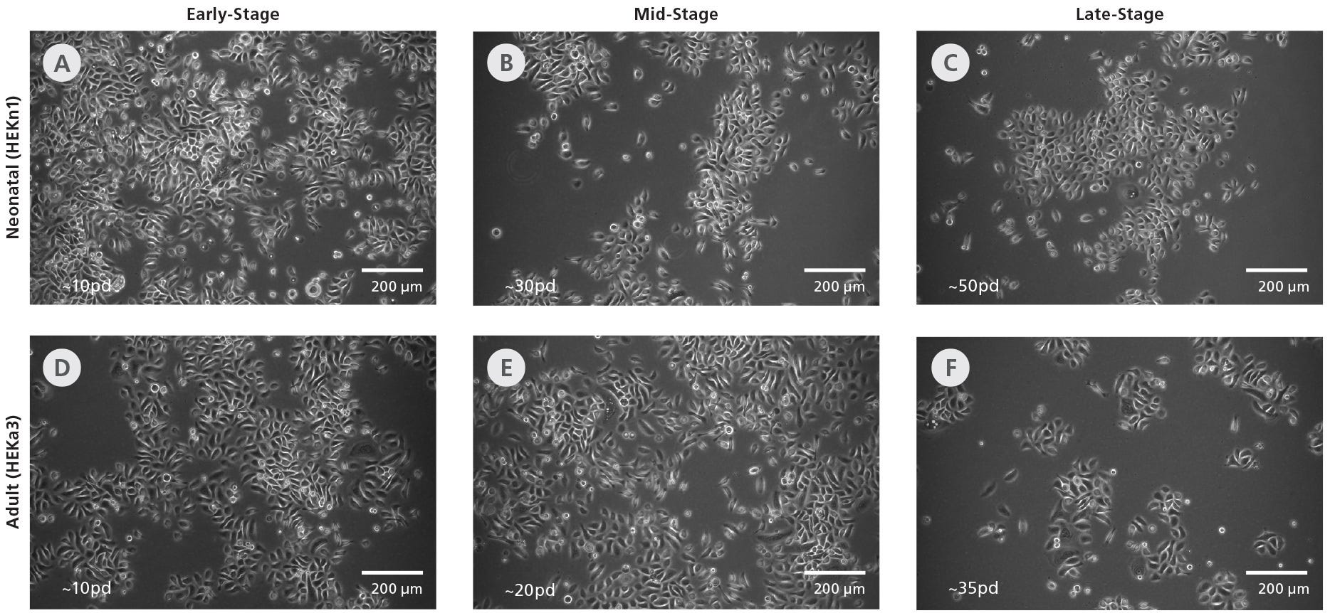
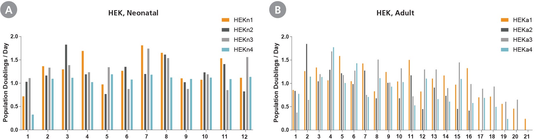
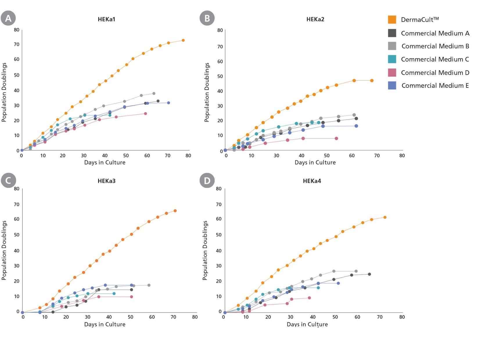
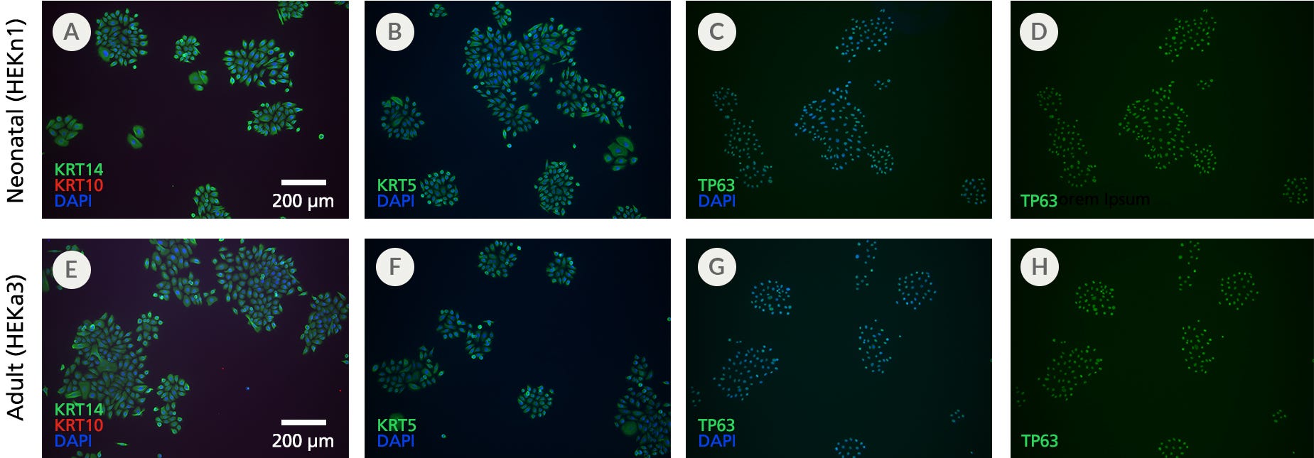
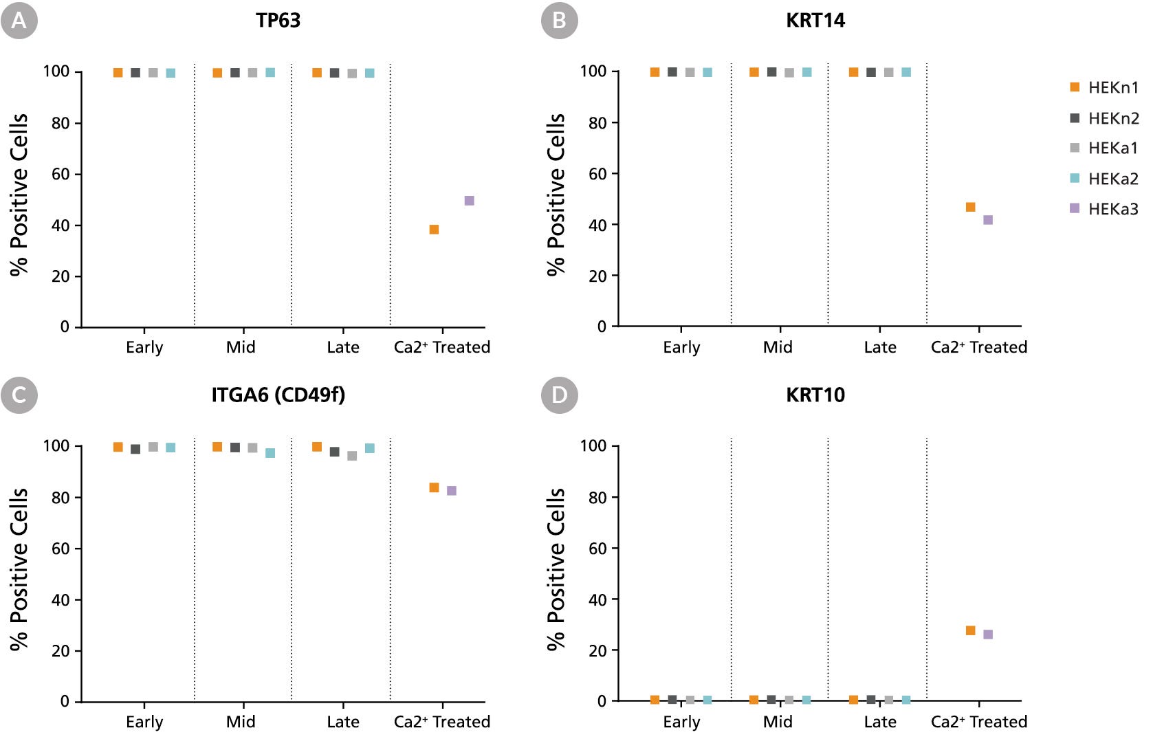
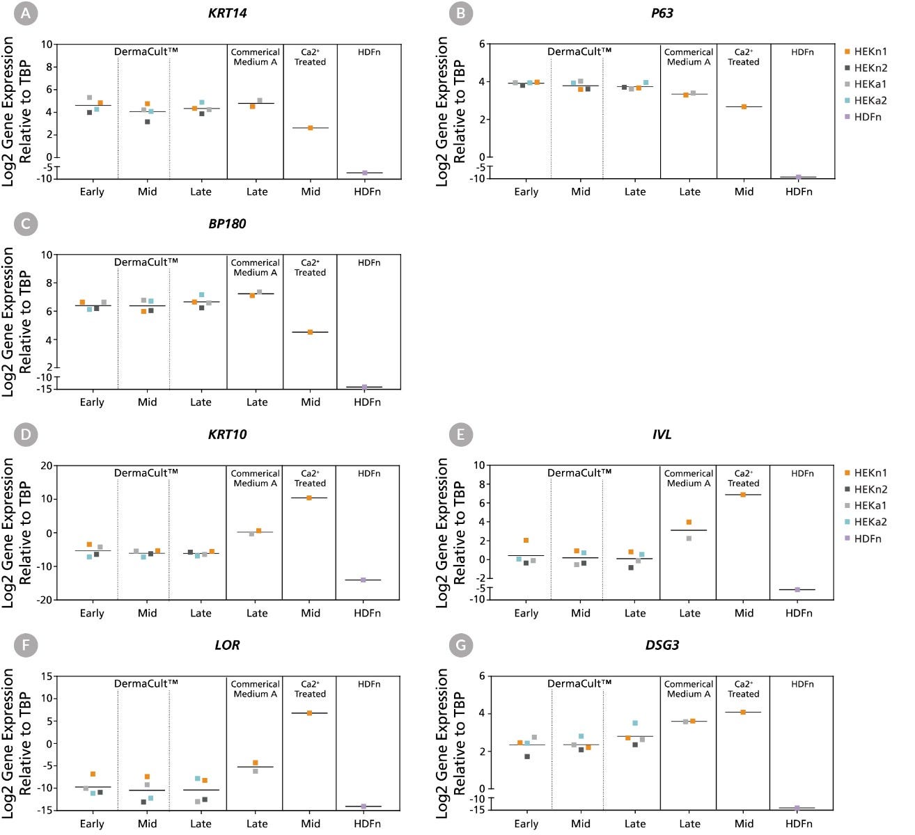
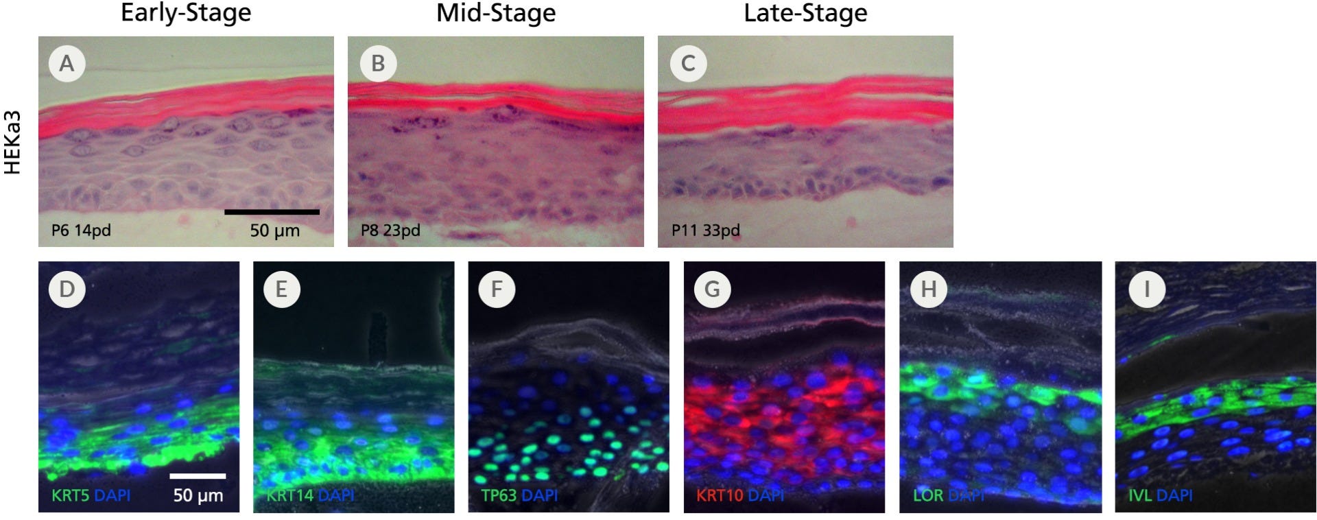
与其他商业培养基不同,DermaCult™ 角质形成细胞扩增培养基可实现人新生儿角质形成细胞超过 50 的群体倍增(population doublings),成人角质形成细胞超过 35 倍增。DermaCult™支持每两天换液一次,并可在任意传代阶段冻存增殖状态的细胞,同时保持其分化潜能,从而方便您开展研究。该培养体系扩增的细胞表现出典型的鹅卵石状形态,并表达代表基底层角质形成细胞的关键标志物(K14、p63、ITGA6 和 ITGB1),可用于后续的气-液界面(ALI)分化培养。详见下方数据。
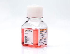
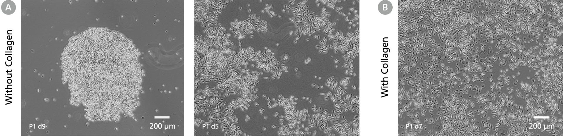
本产品不含血清或 BPE,可最大程度提高实验可重复性。您可以放心地使用在 DermaCult™ 中培养的角质形成细胞作为体外模型,研究皮肤和炎症疾病、进行刺激和腐蚀试验,或评估化妆品或药物的功效。可与 ACCUTASE™(产品号 #07920)搭配使用,推荐每 3 – 5 天传代一次;也适用于胶原蛋白Collagen基质(如:产品号 #04902),例如用于从皮肤组织中建立角质形成细胞培养。
分类
专用培养基,添加剂
细胞类型
真皮细胞,上皮细胞,角质形成细胞
种属
人
应用
细胞培养
品牌
DermaCult
研究领域
癌症,疾病建模,药物发现和毒理检测,移植研究
制剂类别
无血清
请在《产品说明书》中查找相关支持信息和使用说明,或浏览下方更多实验方案。
在线联系


 沪公网安备31010102008431号