若您需要咨询产品或有任何技术问题,请通过官方电话 400 885 9050 或邮箱 info.cn@stemcell.com 与我们联系

DNase I 溶液(1 mg/mL)

细胞解离试剂

产品号 #(选择产品)

产品号 #07900_C

细胞解离试剂

产品优势

  • 使用该即用型溶液,在细胞复苏后可显著减少高浓度或冻存细胞悬液的结块
  • 有效去除解离培养基中的DNA污染物

总览

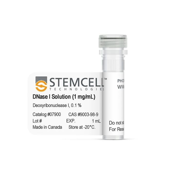
使用脱氧核糖核酸酶(DNase) I溶液(1mg /mL)进行常规组织解离,最大限度地减少在复苏或酶组织消化后高浓度或冻存的细胞悬液的结块,并消除DNA污染物。该即用型溶液的活性范围为每毫克蛋白质≥2000单位。DNase I 由一条带有两个二硫键的糖基化多肽链组成,是一种内切酶,优先切割单链和双链DNA中嘧啶核苷酸附近的磷酸二酯键,生成具有5 ' -磷酸和3 ' -羟基的多核苷酸(Bernardi等)。

包含
• Bovine pancreatic deoxyribonuclease I (1 mg/mL) • Phosphate-buffered saline
 
亚型
酶法相关(或酶解类产品
 
别名
DNA 内切酶;DNA 核酸酶;脱氧核糖核酸磷酸酶;胰 DNase;胸腺核酸酶
 
种属
人,小鼠,非人灵长类,其它细胞系,大鼠
 
应用
细胞培养
 

产品说明书及文档

请在《产品说明书》中查找相关支持信息和使用说明,或浏览下方更多实验方案。

Document Type
Product Name
Catalog #
Lot #
Language
Catalog #
07900, 100-0762
Lot #
All
Language
English
Document Type
Safety Data Sheet
Catalog #
07900
Lot #
All
Language
English

应用领域

本产品专为以下研究领域设计,适用于工作流程中的高亮阶段。探索这些工作流程,了解更多我们为各研究领域提供的其他配套产品。

相关材料与文献

技术资料 (2)

文献 (3)

Intermediate DNA methylation is a conserved signature of genome regulation Elliott G et al. Nature Communications 2015 DEC

Abstract

The role of intermediate methylation states in DNA is unclear. Here, to comprehensively identify regions of intermediate methylation and their quantitative relationship with gene activity, we apply integrative and comparative epigenomics to 25 human primary cell and tissue samples. We report 18,452 intermediate methylation regions located near 36% of genes and enriched at enhancers, exons and DNase I hypersensitivity sites. Intermediate methylation regions average 57% methylation, are predominantly allele-independent and are conserved across individuals and between mouse and human, suggesting a conserved function. These regions have an intermediate level of active chromatin marks and their associated genes have intermediate transcriptional activity. Exonic intermediate methylation correlates with exon inclusion at a level between that of fully methylated and unmethylated exons, highlighting gene context-dependent functions. We conclude that intermediate DNA methylation is a conserved signature of gene regulation and exon usage.
Characterization and ex vivo Expansion of Human Placenta-Derived Natural Killer Cells for Cancer Immunotherapy. Kang L et al. Frontiers in immunology 2013

Abstract

Recent clinical studies suggest that adoptive transfer of donor-derived natural killer (NK) cells may improve clinical outcome in hematological malignancies and some solid tumors by direct anti-tumor effects as well as by reduction of graft versus host disease (GVHD). NK cells have also been shown to enhance transplant engraftment during allogeneic hematopoietic stem cell transplantation (HSCT) for hematological malignancies. The limited ex vivo expansion potential of NK cells from peripheral blood (PB) or umbilical cord blood (UCB) has however restricted their therapeutic potential. Here we define methods to efficiently generate NK cells from donor-matched, full-term human placenta perfusate (termed Human Placenta-Derived Stem Cell, HPDSC) and UCB. Following isolation from cryopreserved donor-matched HPDSC and UCB units, CD56+CD3- placenta-derived NK cells, termed pNK cells, were expanded in culture for up to 3 weeks to yield an average of 1.2 billion cells per donor that were textgreater80% CD56+CD3-, comparable to doses previously utilized in clinical applications. Ex vivo-expanded pNK cells exhibited a marked increase in anti-tumor cytolytic activity coinciding with the significantly increased expression of NKG2D, NKp46, and NKp44 (p textless 0.001, p textless 0.001, and p textless 0.05, respectively). Strong cytolytic activity was observed against a wide range of tumor cell lines in vitro. pNK cells display a distinct microRNA (miRNA) expression profile, immunophenotype, and greater anti-tumor capacity in vitro compared to PB NK cells used in recent clinical trials. With further development, pNK may represent a novel and effective cellular immunotherapy for patients with high clinical needs and few other therapeutic options.
Differential expression of aldehyde dehydrogenase 1a1 (ALDH1) in normal ovary and serous ovarian tumors. Penumatsa K et al. Journal of ovarian research 2010 JAN

Abstract

BACKGROUND: We showed there are specific ALDH1 autoantibodies in ovarian autoimmune disease and ovarian cancer, suggesting a role for ALDH1 in ovarian pathology. However, there is little information on the ovarian expression of ALDH1. Therefore, we compared ALDH1 expression in normal ovary and benign and malignant ovarian tumors to determine if ALDH1 expression is altered in ovarian cancer. Since there is also recent interest in ALDH1 as a cancer stem cell (CSC) marker, we assessed co-expression of ALDH1 with CSC markers in order to determine if ALDH1 is a potential CSC marker in ovarian cancer. METHODS: mRNA and protein expression were compared in normal human ovary and serous ovarian tumors using quantitative Reverse-Transcriptase PCR, Western blot (WB) and semi-quantitative immunohistochemistry (IHC). ALDH1 enzyme activity was confirmed in primary ovarian cells by flow cytometry (FC) using ALDEFLUOR assay. RESULTS: ALDH1 mRNA expression was significantly reduced (p textless 0.01; n = 5) in malignant tumors compared to normal ovaries and benign tumors. The proportion of ALDH1+ cells was significantly lower in malignant tumors (17.1 ± 7.61%; n = 5) compared to normal ovaries (37.4 ± 5.4%; p textless 0.01; n = 5) and benign tumors (31.03 ± 6.68%; p textless 0.05; n = 5). ALDH1+ cells occurred in the stroma and surface epithelium in normal ovary and benign tumors, although surface epithelial expression varied more in benign tumors. Localization of ALDH1 was heterogeneous in malignant tumor cells and little ALDH1 expression occurred in poorly differentiated malignant tumors. In benign tumors the distribution of ALDH1 had features of both normal ovary and malignant tumors. ALDH1 protein expression assessed by IHC, WB and FC was positively correlated (p textless 0.01). ALDH1 did not appear to be co-expressed with the CSC markers CD44, CD117 and CD133 by IHC. CONCLUSIONS: Total ALDH1 expression is significantly reduced in malignant ovarian tumors while it is relatively unchanged in benign tumors compared to normal ovary. Thus, ALDH1 expression in the ovary does not appear to be similar to breast, lung or colon cancer suggesting possible functional differences in these cancers. SIGNIFICANCE: These observations suggest that reduced ALDH1 expression is associated with malignant transformation in ovarian cancer and provides a basis for further study of the mechanism of ALDH1 in this process.

更多信息

更多信息
种属 Human, Mouse, Non-Human Primate, Other, Rat
Contains • Bovine pancreatic deoxyribonuclease I (1 mg/mL) • Phosphate-buffered saline
Alternative Names DNA endonuclease; DNA nuclease; Deoxyribonucleic phosphatase; Pancreatic DNase; Thymonuclease
质量保证:

产品仅供研究使用,不用于针对人或动物的诊断或治疗。 欲获悉更多关于STEMCELL的质控信息,请访问 STEMCELL.CN/COMPLIANCE.
Copyright © 2025 by STEMCELL Technologies. All rights reserved.