
 EasySep™小鼠TIL(CD45)正选试剂盒


产品号 #06030_C
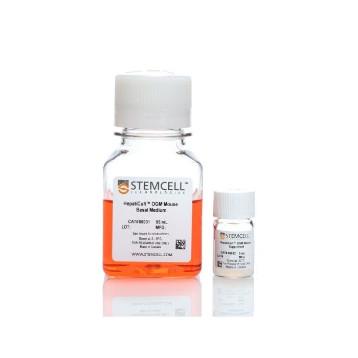
用于建立和维持小鼠肝祖类器官的培养基
若您需要咨询产品或有任何技术问题,请通过官方电话 400 885 9050 或邮箱 info.cn@stemcell.com 与我们联系。
用于建立和维持小鼠肝祖类器官的培养基
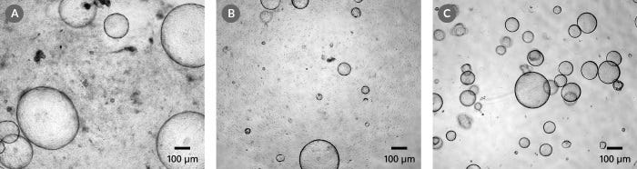
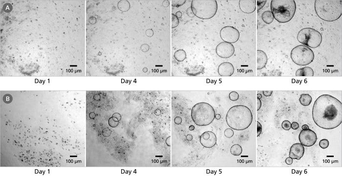
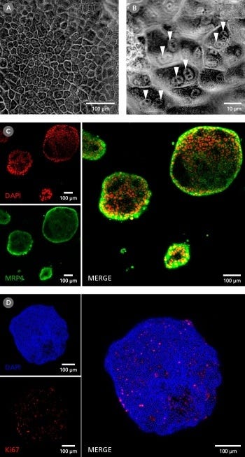
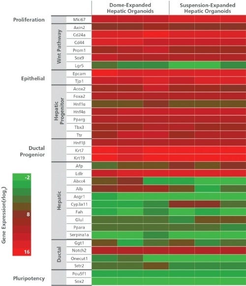
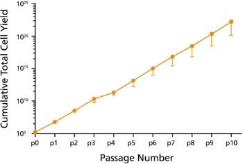
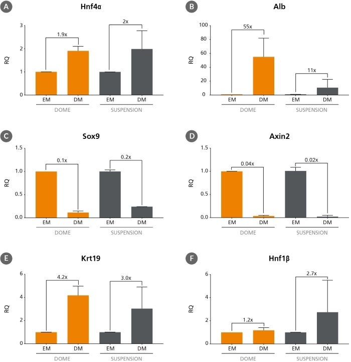
HepatiCult™ 类器官生长培养基 (小鼠)是一款无血清的成分明确的培养基,用于小鼠肝脏祖细胞类器官的建立和维持。这些类器官或称“迷你肝脏”,提供了一个体外的器官型培养系统,用于研究肝脏干细胞和祖细胞。在HepatiCult™培养基中生长的类器官具有表达标记肝脏干细胞和祖细胞(PROM1、AXIN2、SOX9和CD44)、导管细胞(KRT19和HNF1b)以及肝细胞(HNF4a、AFP)基因的上皮。肝类器官可以每4 - 7天传代一次,可进行冷冻保存,也可进行下游分化。
HepatiCult™支持将小鼠肝类器官在Corning® Matrigel®的胶滴中,或在稀释的Matrigel®悬液中进行培养。类器官培养提供了一种便捷的体外方法,可在生理相关的体系中对肝脏上皮进行表征,同时减少对动物的使用。
如果您打算将本产品用于商业目的,请通过www.huborganoids.nl与HUB联系,以获取商业用途许可或HUB许可相关的说明。
分类
专用培养基
细胞类型
肝细胞
种属
小鼠
应用
细胞培养,扩增,培养,类器官培养
品牌
HepatiCult
研究领域
癌症,疾病建模,药物发现和毒理检测,上皮细胞研究,干细胞生物学
制剂类别
无血清
请在《产品说明书》中查找相关支持信息和使用说明,或浏览下方更多实验方案。
本产品专为以下研究领域设计,适用于工作流程中的高亮阶段。探索这些工作流程,了解更多我们为各研究领域提供的其他配套产品。
| 物种 | 小鼠 |
|---|---|
| 配方 | 无血清 |
用于建立和维持小鼠肠道类器官的培养基
用于建立和维持人肠道类器官的培养基
用于人肝脏类器官生成、生长和分化的培养基试剂盒
在线联系


 沪公网安备31010102008431号