
 EasySep™小鼠TIL(CD45)正选试剂盒


产品号 #04962_C
人CFU-Mk染色试剂盒
若您需要咨询产品或有任何技术问题,请通过官方电话 400 885 9050 或邮箱 info.cn@stemcell.com 与我们联系。
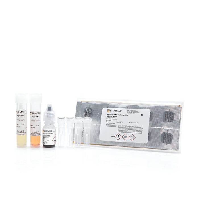
人CFU-Mk染色试剂盒
使用MegaCult™-C CFU-Mk染色试剂盒中包含的试剂,对MegaCult™-C胶原蛋白培养基培养后固定和脱水的CFU-Mk集落进行染色。
•MegaCult™-C一抗
•MegaCult™-C对照抗体
•MegaCult™-C人血清
•MegaCult™-C碱性磷酸酶底物片
•MegaCult™-C生物素偶联山羊抗小鼠IgG
•MegaCult™-C Evans Blue染色剂
•MegaCult™- 10% BSA
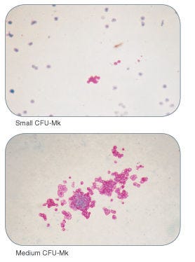
使用抗人CD41抗体和碱性磷酸酶检测系统对表达糖蛋白IIb/IIIa的巨核细胞和血小板进行染色。该试剂盒也适用于检测双潜能红系-巨核细胞祖细胞(BFU-E/Mk)。试剂盒组分可按需单独购买,以方便您的使用。
细胞类型
造血干/祖细胞
种属
人
应用
克隆筛选,免疫细胞化学
品牌
MegaCult
研究领域
干细胞生物学
请在《产品说明书》中查找相关支持信息和使用说明,或浏览下方更多实验方案。
本产品专为以下研究领域设计,适用于工作流程中的高亮阶段。探索这些工作流程,了解更多我们为各研究领域提供的其他配套产品。
| 物种 | 人 |
|---|
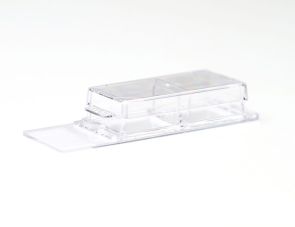
用于胶原基培养物的培养、固定和染色
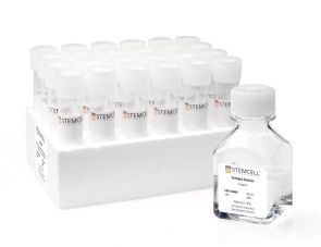
胶原蛋白和不含细胞因子的培养基,用于人和小鼠CFU-Mk检测
胶原蛋白和含细胞因子的培养基,用于人CFU-Mk检测
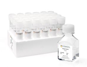
胶原蛋白和含脂质培养基的试剂盒,不含细胞因子,用于人和小鼠CFU-Mk检测
在线联系


 沪公网安备31010102008431号