
 EasySep™小鼠TIL(CD45)正选试剂盒


产品号 #04535_C
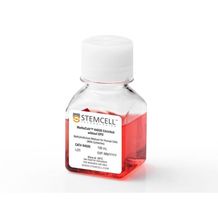
含重组细胞因子(不含EPO)的甲基纤维素培养基,用于人细胞
若您需要咨询产品或有任何技术问题,请通过官方电话 400 885 9050 或邮箱 info.cn@stemcell.com 与我们联系。
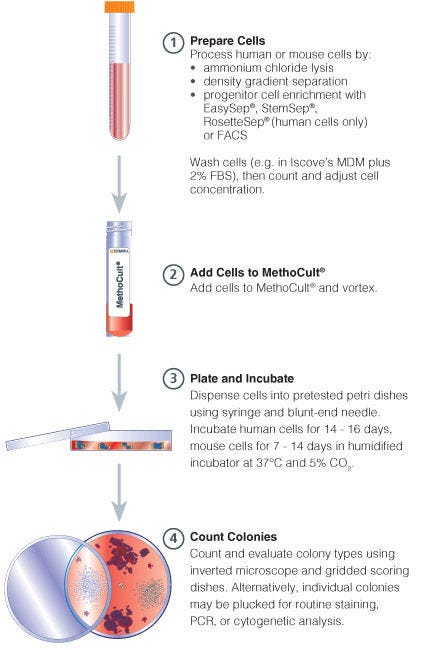
MethoCult™ H4535 Enriched,不含EPO(MethoCult™ GF+ H4535)是一种完全的甲基纤维素培养基,用于人骨髓、动员外周血、外周血和脐带血样本中造血祖细胞在集落形成单位(CFU)检测中的生长和计数。该培养基支持粒细胞-巨噬细胞祖细胞(CFU-GM、CFU-G和CFU-M)的最佳生长,推荐用于CD34+细胞和其他纯化的细胞群体。
参阅关于集落形成单位(CFU)实验的常见问题和解答(FAQ),并探索其作为细胞治疗工作流程组成部分的应用价值。
包含
• 含甲基纤维素的Iscove's MDM
• 胎牛血清
• 牛血清白蛋白
• 2-巯基乙醇
• 重组人干细胞因子 (SCF)
• 重组人白细胞介素3 (IL-3)
• 重组人白细胞介素6 (IL-6)
• 重组人粒细胞集落刺激因子 (G-CSF)
• 重组人粒细胞-巨噬细胞集落刺激因子 (GM-CSF)
• 添加物
分类
半固体培养基,专用培养基
细胞类型
造血干/祖细胞
种属
人、非人灵长类
应用
细胞培养,克隆筛选,功能学筛选
品牌
MethoCult
研究领域
药物发现和毒性检测,干细胞生物学
请在《产品说明书》中查找相关支持信息和使用说明,或浏览下方更多实验方案。
本产品专为以下研究领域设计,适用于工作流程中的高亮阶段。探索这些工作流程,了解更多我们为各研究领域提供的其他配套产品。
| 物种 | 人, 非人灵长类 |
|---|---|
| Contains | • Methylcellulose in Iscove's MDM • Fetal bovine serum • Bovine serum albumin • 2-Mercaptoethanol • Recombinant human stem cell factor (SCF) • Recombinant human interleukin 3 (IL-3) • Recombinant human interleukin 6 (IL-6) • Recombinant human granulocyte |
用于人细胞的含重组细胞因子的甲基纤维素培养基
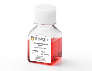
用于制备和洗涤CFU检测样本的培养基
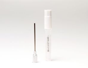
16号鲁尔锁针头
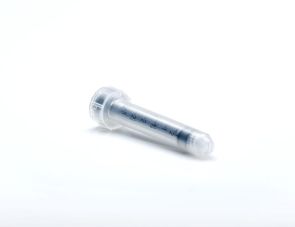
用于精确分装甲基纤维素培养基
在线联系


 沪公网安备31010102008431号