
 EasySep™小鼠TIL(CD45)正选试剂盒


产品号 #05982
用于人骨骼肌祖细胞(成肌细胞)衍生和扩增的无血清添加物
若您需要咨询产品或有任何技术问题,请通过官方电话 400 885 9050 或邮箱 info.cn@stemcell.com 与我们联系。
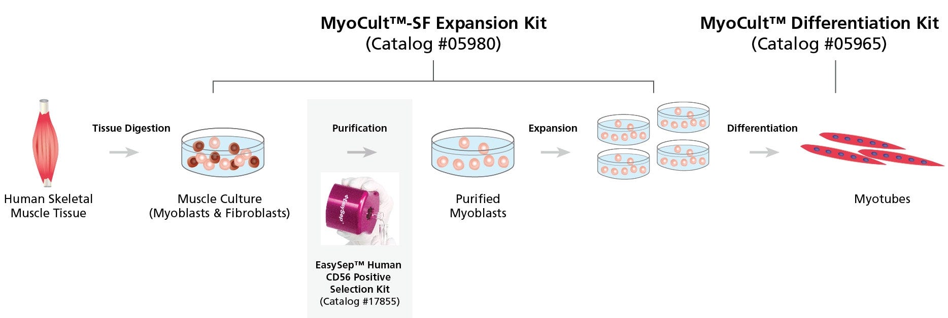
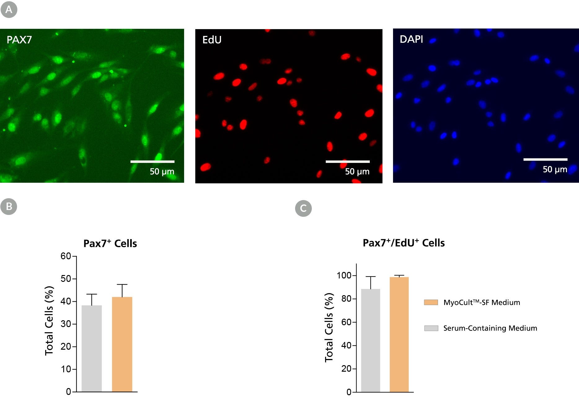
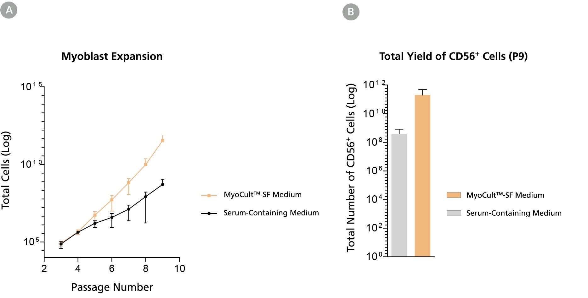
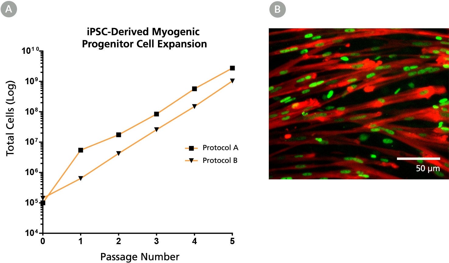
使用 MyoCult™-SF 扩增10X添加物(人)支持原代人骨骼肌祖细胞和人多能干细胞(hPSC)来源成肌细胞的无血清培养。
该补充剂可单独购买或作为 MyoCult™-SF 扩增添加物试剂盒 (人) 的一部分提供,采用无血清配方,必须与基础培养基(含 1000 mg/L D-葡萄糖的 DMEM(产品号 #36253);单独销售)混合以配制 MyoCult™-SF 扩增培养基。该培养基经过优化,适用于衍生和体外扩增人骨骼肌祖细胞。
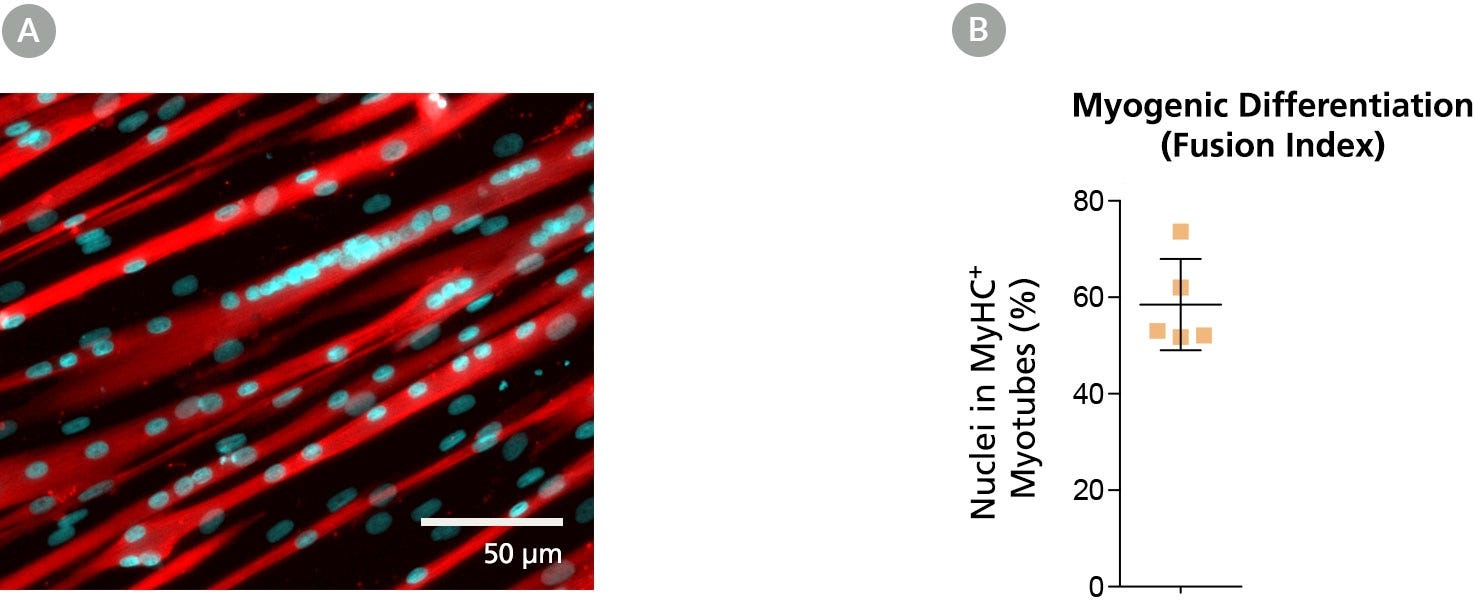
若使用原代成肌细胞,建议用MyoCult™-SF 贴附基质包被培养器皿。对于 hPSC 来源的成肌细胞,可使用 Corning® Matrigel® hESC-Qualified Matrix。使用 MyoCult™-SF 扩增培养基培养的成肌细胞与 MyoCult™ 分化试剂盒(人)兼容,可用于进一步分化为多核肌管结构。
有关使用 MyoCult™ 进行人骨骼肌祖细胞衍生和扩增的详细方案,请参阅产品说明书。
分类
专用培养基
细胞类型
肌源干/祖细胞
种属
人
应用
细胞培养,扩增
品牌
MyoCult
研究领域
干细胞生物学
制剂类别
无血清
请在《产品说明书》中查找相关支持信息和使用说明,或浏览下方更多实验方案。
本产品专为以下研究领域设计,适用于工作流程中的高亮阶段。探索这些工作流程,了解更多我们为各研究领域提供的其他配套产品。
| 物种 | 人 |
|---|---|
| 配方 | 无血清 |
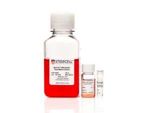
人骨骼肌祖细胞(成肌细胞)分化为肌管的培养基
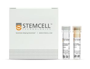
人CD56+细胞的免疫磁珠正选
小鼠Monoclonal IgG1抗体,抗人、黑猩猩CD45
小鼠Monoclonal IgG1抗体,抗人CD56 (NCAM)
在线联系


 沪公网安备31010102008431号