
 EasySep™小鼠TIL(CD45)正选试剂盒


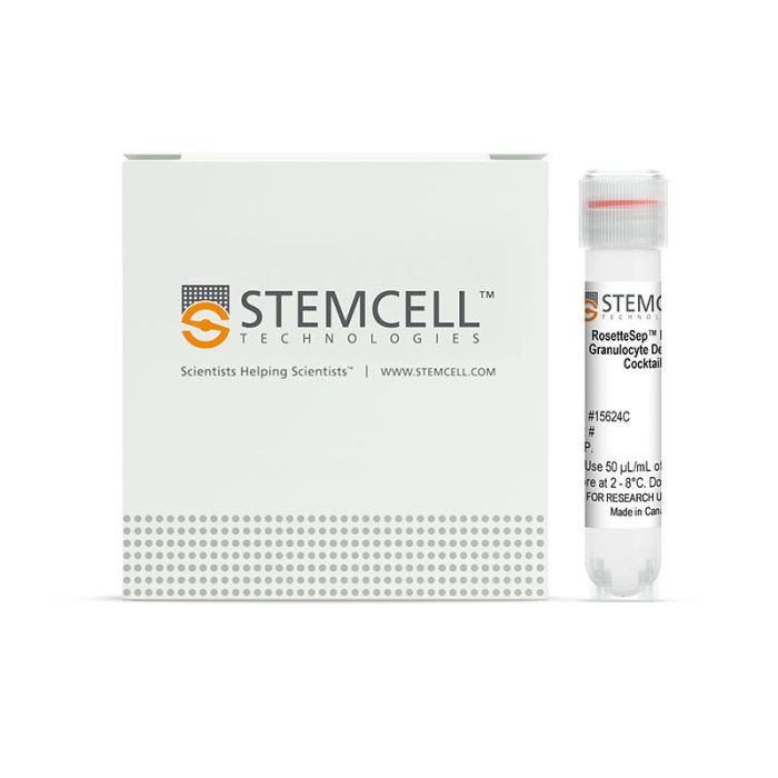
产品号 #15624_C
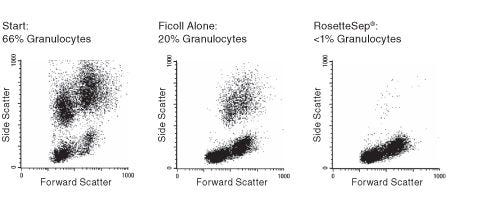
免疫密度梯度离心法去除粒细胞
若您需要咨询产品或有任何技术问题,请通过官方电话 400 885 9050 或邮箱 info.cn@stemcell.com 与我们联系。
免疫密度梯度离心法去除粒细胞
RosetteSep™ 人粒细胞去除复合物专门为通过负选从全血中去除粒细胞而设计。利用识别 CD66b 与红细胞膜糖蛋白 A 的四聚抗体复合物标记想去除的粒细胞,在 Lymphoprep™(产品号#18060)上离心后,标记细胞与红细胞共同沉淀在底部,去除了粒细胞的细胞群位于血浆与密度介质交界面。
分类
细胞分选试剂盒
细胞类型
粒细胞及其亚群
种属
人
样本来源
白膜层、全血
分选方法
去除
应用
细胞分选
品牌
RosetteSep
研究领域
免疫
请在《产品说明书》中查找相关支持信息和使用说明,或浏览下方更多实验方案。
本产品专为以下研究领域设计,适用于工作流程中的高亮阶段。探索这些工作流程,了解更多我们为各研究领域提供的其他配套产品。
| 物种 | 人 |
|---|---|
| 样本来源 | 全血, 白膜层 |
| Selection Method | Depletion |
直接从全血中免疫磁珠分选中性粒细胞
用于体外诊断(IVD)应用的密度梯度离心管
在线联系


 沪公网安备31010102008431号