Biophysical regulation of epigenetic state and cell reprogramming
Biochemical factors can help reprogram somatic cells into pluripotent stem cells,yet the role of biophysical factors during reprogramming is unknown. Here,we show that biophysical cues,in the form of parallel microgrooves on the surface of cell-adhesive substrates,can replace the effects of small-molecule epigenetic modifiers and significantly improve reprogramming efficiency. The mechanism relies on the mechanomodulation of the cells' epigenetic state. Specifically,decreased histone deacetylase activity and upregulation of the expression of WD repeat domain 5 (WDR5)—a subunit of H3 methyltranferase—by microgrooved surfaces lead to increased histone H3 acetylation and methylation. We also show that microtopography promotes a mesenchymal-to-epithelial transition in adult fibroblasts. Nanofibrous scaffolds with aligned fibre orientation produce effects similar to those produced by microgrooves,suggesting that changes in cell morphology may be responsible for modulation of the epigenetic state. These findings have important implications in cell biology and in the optimization of biomaterials for cell-engineering applications.
View Publication
产品号#:
05850
05857
05870
05875
85850
85857
85870
85875
产品名:
mTeSR™1
mTeSR™1
Cheng Y et al. ( 2013)
BMC cell biology 14 1 44
Physiological β-catenin signaling controls self-renewal networks and generation of stem-like cells from nasopharyngeal carcinoma.
BACKGROUND: A few reports suggested that low levels of Wnt signaling might drive cell reprogramming,but these studies could not establish a clear relationship between Wnt signaling and self-renewal networks. There are ongoing debates as to whether and how the Wnt/β-catenin signaling is involved in the control of pluripotency gene networks. Additionally,whether physiological β-catenin signaling generates stem-like cells through interactions with other pathways is as yet unclear. The nasopharyngeal carcinoma HONE1 cells have low expression of β-catenin and wild-type expression of p53,which provided a possibility to study regulatory mechanism of stemness networks induced by physiological levels of Wnt signaling in these cells.backslashnbackslashnRESULTS: Introduction of increased β-catenin signaling,haploid expression of β-catenin under control by its natural regulators in transferred chromosome 3,resulted in activation of Wnt/β-catenin networks and dedifferentiation in HONE1 hybrid cell lines,but not in esophageal carcinoma SLMT1 hybrid cells that had high levels of endogenous β-catenin expression. HONE1 hybrid cells displayed stem cell-like properties,including enhancement of CD24(+) and CD44(+) populations and generation of spheres that were not observed in parental HONE1 cells. Signaling cascades were detected in HONE1 hybrid cells,including activation of p53- and RB1-mediated tumor suppressor pathways,up-regulation of Nanog-,Oct4-,Sox2-,and Klf4-mediated pluripotency networks,and altered E-cadherin expression in both in vitro and in vivo assays. qPCR array analyses further revealed interactions of physiological Wnt/β-catenin signaling with other pathways such as epithelial-mesenchymal transition,TGF-β,Activin,BMPR,FGFR2,and LIFR- and IL6ST-mediated cell self-renewal networks. Using β-catenin shRNA inhibitory assays,a dominant role for β-catenin in these cellular network activities was observed. The expression of cell surface markers such as CD9,CD24,CD44,CD90,and CD133 in generated spheres was progressively up-regulated compared to HONE1 hybrid cells. Thirty-four up-regulated components of the Wnt pathway were identified in these spheres.backslashnbackslashnCONCLUSIONS: Wnt/β-catenin signaling regulates self-renewal networks and plays a central role in the control of pluripotency genes,tumor suppressive pathways and expression of cancer stem cell markers. This current study provides a novel platform to investigate the interaction of physiological Wnt/β-catenin signaling with stemness transition networks.
View Publication
Yamashita J et al. (NOV 2000)
Nature 408 6808 92--6
Flk1-positive cells derived from embryonic stem cells serve as vascular progenitors.
Interaction between endothelial cells and mural cells (pericytes and vascular smooth muscle) is essential for vascular development and maintenance. Endothelial cells arise from Flk1-expressing (Flk1+) mesoderm cells,whereas mural cells are believed to derive from mesoderm,neural crest or epicardial cells and migrate to form the vessel wall. Difficulty in preparing pure populations of these lineages has hampered dissection of the mechanisms underlying vascular formation. Here we show that Flk1+ cells derived from embryonic stem cells can differentiate into both endothelial and mural cells and can reproduce the vascular organization process. Vascular endothelial growth factor promotes endothelial cell differentiation,whereas mural cells are induced by platelet-derived growth factor-BB. Vascular cells derived from Flk1+ cells can organize into vessel-like structures consisting of endothelial tubes supported by mural cells in three-dimensional culture. Injection of Flk1+ cells into chick embryos showed that they can incorporate as endothelial and mural cells and contribute to the developing vasculature in vivo. Our findings indicate that Flk1+ cells can act as 'vascular progenitor cells' to form mature vessels and thus offer potential for tissue engineering of the vascular system.
View Publication

 EasySep™小鼠TIL(CD45)正选试剂盒


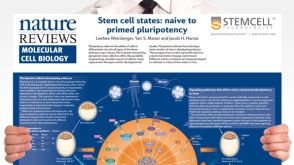 挂图Stem Cell States: Naive to Primed Pluripotency Properties of naive (ground) and primed pluripotent stem cells
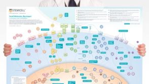 挂图Small Molecules, Big Impact in Pluripotent Stem Cell Research Overview of signaling pathways and small molecules in pluripotent stem cell research
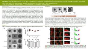 科学海报STEMdiff™ Cerebral Organoid Kit: A New Tool for the Culture of 3D Brain Organoids Derived from hPSCs


 沪公网安备31010102008431号