
 EasySep™小鼠TIL(CD45)正选试剂盒


技术资料
-

-
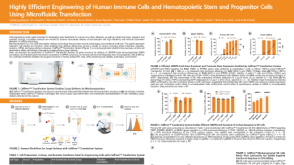
 实验方案How to Prepare and Plate Semi-Solid Methylcellulose Medium for Cell Culture
研究方向:
干细胞生物学,细胞系制备,药物发现和毒理检测
-

-

-

-


 沪公网安备31010102008431号