
 EasySep™小鼠TIL(CD45)正选试剂盒


技术资料
-
产品号#:
05835
05839
产品名:
STEMdiff™ 神经诱导培养基
STEMdiff™ 神经诱导培养基
-
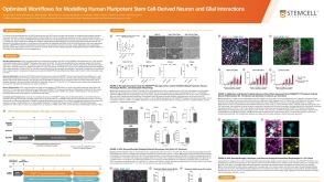 科学海报Optimized Workflows for Modelling Human Pluripotent Stem Cell-Derived Neuron and Glial Interactions
Conference:
Society for Neuroscience (SfN)
-
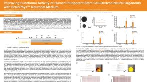 科学海报Improving Functional Activity of Human Pluripotent Stem Cell-Derived Neural Organoids with BrainPhys Neuronal Medium
Conference:
Society for Neuroscience (SfN)
-
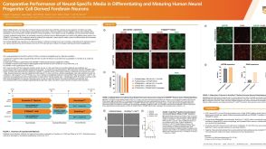 科学海报Comparative Performance of Neural-Specific Media in Differentiating and Maturing Human Neural Progenitor Cell-Derived Forebrain Neurons
Conference:
Society for Neuroscience (SfN)
-
 实验方案How to Generate AssemBloids™ from hPSC-Derived Dorsal and Ventral Forebrain Organoid Co-Cultures
研究方向:
疾病建模,神经科学,类器官,药物发现和毒性检测


 沪公网安备31010102008431号