A. Holtzinger et al. ( 2015)
Development (Cambridge,England) 142 4253-65
New markers for tracking endoderm induction and hepatocyte differentiation from human pluripotent stem cells.
The efficient generation of hepatocytes from human pluripotent stem cells (hPSCs) requires the induction of a proper endoderm population,broadly characterized by the expression of the cell surface marker CXCR4. Strategies to identify and isolate endoderm subpopulations predisposed to the liver fate do not exist. In this study,we generated mouse monoclonal antibodies against human embryonic stem cell-derived definitive endoderm with the goal of identifying cell surface markers that can be used to track the development of this germ layer and its specification to a hepatic fate. Through this approach,we identified two endoderm-specific antibodies,HDE1 and HDE2,which stain different stages of endoderm development and distinct derivative cell types. HDE1 marks a definitive endoderm population with high hepatic potential,whereas staining of HDE2 tracks with developing hepatocyte progenitors and hepatocytes. When used in combination,the staining patterns of these antibodies enable one to optimize endoderm induction and hepatic specification from any hPSC line.
View Publication
Řeboun M et al. ( 2016)
Folia biologica 62 2 82--89
X-Chromosome Inactivation Analysis in Different Cell Types and Induced Pluripotent Stem Cells Elucidates the Disease Mechanism in a Rare Case of Mucopolysaccharidosis Type II in a Female.
Mucopolysaccharidosis type II (MPS II) is an X-linked lysosomal storage disorder resulting from deficiency of iduronate-2-sulphatase activity. The disease manifests almost exclusively in males; only 16 symptomatic heterozygote girls have been reported so far. We describe the results of X-chromosome inactivation analysis in a 5-year-old girl with clinically severe disease and heterozygous mutation p.Arg468Gln in the IDS gene. X inactivation analysed at three X-chromosome loci showed extreme skewing (96/4 to 99/1) in two patient's cell types. This finding correlated with exclusive expression of the mutated allele. Induced pluripotent stem cells (iPSC) generated from the patient's peripheral blood demonstrated characteristic pluripotency markers,deficiency of enzyme activity,and mutation in the IDS gene. These cells were capable of differentiation into other cell types (cardiomyocytes,neurons). In MPS II iPSC clones,the X inactivation ratio remained highly skewed in culture conditions that led to partial X inactivation reset in Fabry disease iPSC clones. Our data,in accordance with the literature,suggest that extremely skewed X inactivation favouring the mutated allele is a crucial condition for manifestation of MPS II in females. This suggests that the X inactivation status and enzyme activity have a prognostic value and should be used to evaluate MPS II in females. For the first time,we show generation of iPSC from a symptomatic MPS II female patient that can serve as a cellular model for further research of the pathogenesis and treatment of this disease.
View Publication
Non-integrating episomal plasmid-based reprogramming of human amniotic fluid stem cells into induced pluripotent stem cells in chemically defined conditions.
Amniotic fluid stem cells (AFSC) represent an attractive potential cell source for fetal and pediatric cell-based therapies. However,upgrading them to pluripotency confers refractoriness toward senescence,higher proliferation rate and unlimited differentiation potential. AFSC were observed to rapidly and efficiently reacquire pluripotency which together with their easy recovery makes them an attractive cell source for reprogramming. The reprogramming process as well as the resulting iPSC epigenome could potentially benefit from the unspecialized nature of AFSC. iPSC derived from AFSC also have potential in disease modeling,such as Down syndrome or $\$-thalassemia. Previous experiments involving AFSC reprogramming have largely relied on integrative vector transgene delivery and undefined serum-containing,feeder-dependent culture. Here,we describe non-integrative oriP/EBNA-1 episomal plasmid-based reprogramming of AFSC into iPSC and culture in fully chemically defined xeno-free conditions represented by vitronectin coating and E8 medium,a system that we found uniquely suited for this purpose. The derived AF-iPSC lines uniformly expressed a set of pluripotency markers Oct3/4,Nanog,Sox2,SSEA-1,SSEA-4,TRA-1-60,TRA-1-81 in a pattern typical for human primed PSC. Additionally,the cells formed teratomas,and were deemed pluripotent by PluriTest,a global expression microarray-based in-silico pluripotency assay. However,we found that the PluriTest scores were borderline,indicating a unique pluripotent signature in the defined condition. In the light of potential future clinical translation of iPSC technology,non-integrating reprogramming and chemically defined culture are more acceptable.
View Publication
产品号#:
05850
05857
05870
05875
05940
07180
07183
07190
27147
07191
07930
07931
07940
07955
07956
07959
07954
85850
85857
85870
85875
100-1061
07952
100-0763
产品名:
Vitronectin XF™
CellAdhere™ 稀释缓冲液
CryoStor® CS10
CryoStor® CS10
CryoStor® CS10
CryoStor® CS10
CryoStor® CS10
mTeSR™1
mTeSR™1
CryoStor® CS10
CryoStor® CS10
Vitronectin XF™
Koh S and Piedrahita JA ( 2015)
1330 69--78
Generation of induced pluripotent stem cells (iPSCs) from adult canine fibroblasts
Induced pluripotent stem cells hold great potential in regenerative medicine as it enables to generate pluripotent stem cells from any available cell types. Ectopic expression of four transcription factors (Oct4,Sox2,Klf4,and c-Myc) can reprogram fibroblasts directly to pluripotency as shown in multiple species. Here,we describe detailed protocols for generation of iPSCs from adult canine fibroblasts. Robust canine iPSCs will provide powerful tools not only to study human diseases,but also for the development of therapeutic approaches.
View Publication
产品号#:
05850
05857
05870
05875
85850
85857
85870
85875
产品名:
mTeSR™1
mTeSR™1
Lee Y-LL et al. (NOV 2015)
Human reproduction (Oxford,England) 30 11 2614--2626
Establishment of a novel human embryonic stem cell-derived trophoblastic spheroid implantation model.
STUDY QUESTION Can human embryonic stem cell-derived trophoblastic spheroids be used to study the early stages of implantation? SUMMARY ANSWER We generated a novel human embryonic stem cell-derived trophoblastic spheroid model mimicking human blastocysts in the early stages of implantation. WHAT IS KNOWN ALREADY Both human embryos and choriocarcinoma cell line derived spheroids can attach onto endometrial cells and are used as models to study the early stages of implantation. However,human embryos are limited and the use of cancer cell lines for spheroid generation remains sub-optimal for research. STUDY DESIGN,SIZE,DURATION Experimental induced differentiation of human embryonic stem cells into trophoblast and characterization of the trophoblast. PARTICIPANTS/MATERIALS,SETTING,METHODS Trophoblastic spheroids (BAP-EB) were generated by inducing differentiation of a human embryonic stem cell line,VAL3 cells with bone morphogenic factor-4,A83-01 (a TGF-$\$),and PD173074 (a FGF receptor-3 inhibitor) after embryoid body formation. The expressions of trophoblastic markers and hCG levels were studied by real-time PCR and immunohistochemistry. BAP-EB attachment and invasion assays were performed on different cell lines and primary endometrial cells. MAIN RESULTS AND THE ROLE OF CHANCE After 48 h of induced differentiation,the BAP-EB resembled early implanting human embryos in terms of size and morphology. The spheroids derived from embryonic stem cells (VAL3),but not from several other cell lines studied,possessed a blastocoel-like cavity. BAP-EB expressed several markers of trophectoderm of human blastocysts on Day 2 of induced differentiation. In the subsequent days of differentiation,the cells of the spheroids differentiated into trophoblast-like cells expressing trophoblastic markers,though at levels lower than that in the primary trophoblasts or in a choriocarcinoma cell line. On Day 3 of induced differentiation,BAP-EB selectively attached onto endometrial epithelial cells,but not other non-endometrial cell lines or an endometrial cell line that had lost its epithelial character. The attachment rates of BAP-EB was significantly higher on primary endometrial epithelial cells (EEC) taken from 7 days after hCG induction of ovulation (hCG+7 day) when compared with that from hCG+2 day. The spheroids also invaded through Ishikawa cells and the primary endometrial stromal cells in the co-culture. LIMITATIONS,REASONS FOR CAUTION The attachment rates of BAP-EB were compared between EEC obtained from Day 2 and Day 7 of the gonadotrophin stimulated cycle,but not the natural cycles. WIDER IMPLICATIONS OF THE FINDINGS BAP-EB have the potential to be used as a test for predicting endometrial receptivity in IVF cycles and provide a novel approach to study early human implantation,trophoblastic cell differentiation and trophoblastic invasion into human endometrial cells.
View Publication

 EasySep™小鼠TIL(CD45)正选试剂盒


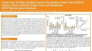 科学海报Expansion of High-Quality Human Pluripotent Stem Cells (hPSCs) Using a Novel Animal Origin-Free and Stabilized hPSC Maintenance Medium
 实验方案Large-Scale Expansion of hPSCs in 2D Monolayer Culture using mTeSR™ Plus
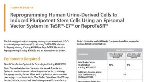


 沪公网安备31010102008431号