The MADS transcription factor Mef2c is a pivotal modulator of myeloid cell fate.
Mef2c is a MADS (MCM1-agamous-deficient serum response factor) transcription factor best known for its role in muscle and cardiovascular development. A causal role of up-regulated MEF2C expression in myelomonocytic acute myeloid leukemia (AML) has recently been demonstrated. Due to the pronounced monocytic component observed in Mef2c-induced AML,this study was designed to assess the importance of Mef2c in normal myeloid differentiation. Analysis of bone marrow (BM) cells manipulated to constitutively express Mef2c demonstrated increased monopoiesis at the expense of granulopoiesis,whereas BM isolated from Mef2c(Delta/-) mice showed reduced levels of monocytic differentiation in response to cytokines. Mechanistic studies showed that loss of Mef2c expression correlated with reduced levels of transcripts encoding c-Jun,but not PU.1,C/EBPalpha,or JunB transcription factors. Inhibiting Jun expression by short-interfering RNA impaired Mef2c-mediated inhibition of granulocyte development. Moreover,retroviral expression of c-Jun in BM cells promoted monocytic differentiation. The ability of Mef2c to modulate cell-fate decisions between monocyte and granulocyte differentiation,coupled with its functional sensitivity to extracellular stimuli,demonstrate an important role in immunity--and,consistent with findings of other myeloid transcription factors,a target of oncogenic lesions in AML.
View Publication
产品号#:
03434
03444
09600
09650
18556
18556RF
产品名:
MethoCult™ GF M3434
MethoCult™ GF M3434
StemSpan™ SFEM
StemSpan™ SFEM
Soto-Cruz I et al. ( 2008)
Cancer Investigation 26 2 136--144
The Tyrphostin B42 Inhibits Cell Proliferation and HER-2 Autophosphorylation in Cervical Carcinoma Cell Lines
The HER family receptors have an important role controlling cell growth and differentiation. Although the activity of the HER-2 receptor is strictly controlled in normal cells,its overexpression plays a pivotal role in transformation and tumorigenesis. Constitutive phosphorylation of HER-2 protein has been implicated in conferring uncontrolled growth to mammary cancer cells,and to a lesser extent,with adenocarcinoma of uterus,cervix,fallopian tube,and endometrium. This study addresses the role of HER-2 in cervical carcinoma. Firstly,we demonstrate the presence of HER-2 protein expression by flow cytometry in two new cervical carcinoma cell lines CALO and INBL. Secondly,we use the specific tyrosine kinase inhibitors,Tyrphostins to examine HER-2 regulation by the crystal violet assay. Thirdly,we use western blot analysis to assess the state of HER-2 phosphorylation. The most efficient agent,Tyrphostin B42,known as an inhibitor of epithelial growth factor receptor,arrested cervical carcinoma cell lines growth in vitro at micromolar concentrations within 72 h of application. Tyrphostin B42 inhibited the HER2 signal-regulated kinase pathway,as observed by the reduction in the phosphorylated forms of HER2. The loss of phosphorylated forms of HER2 at early time points after Tyrphostin B42 application was associated with suppression of cell growth. Thus,the inhibition of the proliferation of our cervical carcinoma cell lines by Tyrphostin B42 is associated with inhibition of HER2 protein kinase signal.
View Publication
产品号#:
72932
72934
产品名:
AG - 490
Brandl M et al. (AUG 1999)
Experimental hematology 27 8 1264--70
Bispecific antibody fragments with CD20 X CD28 specificity allow effective autologous and allogeneic T-cell activation against malignant cells in peripheral blood and bone marrow cultures from patients with B-cell lineage leukemia and lymphoma.
Bispecific antibodies directed against tumor-associated target antigens and to surface receptors mediating T-cell activation,such as the TCR/CD3 complex and the costimulatory receptor CD28,are capable of mediating T-cell activation resulting in tumor cell killing. In this study,we used the B-cell-associated antigens CD19 and CD20 as target structures on human leukemic cells. We found that a combination of bispecific antibody fragments (bsFab2) with target x CD3 and target x CD28 specificity induces vigorous autologous T-cell activation and killing of malignant cells in peripheral blood and bone marrow cultures from patients with chronic lymphocytic leukemia and follicular lymphoma. The bsFab2 targeting CD20 were considerably more effective than those binding to CD19. The colony-forming capacity of treated bone marrow was impaired due to large amounts of tumor necrosis factor alpha produced during bsFab2-induced T-cell activation. Neutralizing tumor necrosis factor alpha antibodies were found to reverse this negative effect without affecting T-cell activation and tumor cell killing. CD20 x CD28 bsFab2,when used alone rather than in combination,markedly improved the recognition of leukemic cells by allogeneic T cells. Therefore,these reagents may be capable of enhancing the immunogenicity of leukemic cells in general and,in particular,of increasing the antileukemic activity of allogeneic donor buffy coat cells in relapsed bone marrow transplanted patients.
View Publication
Liu Z et al. (FEB 2012)
Journal of stem cell research & therapy 2 1 1--8
Blockade of Autocrine TGF-$$ Signaling Inhibits Stem Cell Phenotype, Survival, and Metastasis of Murine Breast Cancer Cells.
Transforming growth factor beta (TGF-$$) signaling has been implicated in driving tumor progression and metastasis by inducing stem cell-like features in some human cancer cell lines. In this study,we have utilized a novel murine cell line NMuMG-ST,which acquired cancer stem cell (CSC) phenotypes during spontaneous transformation of the untransformed murine mammary cell line NMuMG,to investigate the role of autocrine TGF-$$ signaling in regulating their survival,metastatic ability,and the maintenance of cancer stem cell characteristics. We have retrovirally transduced a dominant-negative TGF-$$ type II receptor (DNRII) into the NMuMG-ST cell to abrogate autocrine TGF-$$ signaling. The expression of DNRII reduced TGF-$$ sensitivity of the NMuMG-ST cells in various cell-based assays. The blockade of autocrine TGF-$$ signaling reduced the ability of the cell to grow anchorage-independently and to resist serum deprivation-induced apoptosis. These phenotypes were associated with reduced levels of active and phosphorylated AKT and ERK,and Gli1 expression suggesting that these pathways contribute to the growth and survival of this model system. More interestingly,the abrogation of autocrine TGF-$$ signaling also led to the attenuation of several features associated with mammary stem cells including epithelial-mesenchymal transition,mammosphere formation,and expression of stem cell markers. When xenografted in athymic nude mice,the DNRII cells were also found to undergo apoptosis and induced significantly lower lung metastasis burden than the control cells even though they formed similar size of xenograft tumors. Thus,our results indicate that autocrine TGF-$$ signaling is involved in the maintenance and survival of stem-like cell population resulting in the enhanced metastatic ability of the murine breast cancer cells.
View Publication

 EasySep™小鼠TIL(CD45)正选试剂盒


 挂图Natural Killer Cells Overview of NK cell receptors, subsets, activation and function
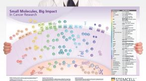 挂图Small Molecules, Big Impact in Cancer Research Overview of signaling pathways and small molecules in cancer research
 挂图Small Molecules, Big Impact in Pluripotent Stem Cell Research Overview of signaling pathways and small molecules in pluripotent stem cell research

 沪公网安备31010102008431号