Li Y et al. (JAN 2016)
Journal of virology 90 7 3385--99
Ecotropic Murine Leukemia Virus Infection of Glial Progenitors Interferes with Oligodendrocyte Differentiation: Implications for Neurovirulence.
UNLABELLED Certain murine leukemia viruses (MLVs) are capable of inducing fatal progressive spongiform motor neuron disease in mice that is largely mediated by viral Env glycoprotein expression within central nervous system (CNS) glia. While the etiologic mechanisms and the glial subtypes involved remain unresolved,infection of NG2 glia was recently observed to correlate spatially and temporally with altered neuronal physiology and spongiogenesis. Since one role of NG2 cells is to serve as oligodendrocyte (OL) progenitor cells (OPCs),we examined here whether their infection by neurovirulent (FrCasE) or nonneurovirulent (Fr57E) ecotropic MLVs influenced their viability and/or differentiation. Here,we demonstrate that OPCs,but not OLs,are major CNS targets of both FrCasE and Fr57E. We also show that MLV infection of neural progenitor cells (NPCs) in culture did not affect survival,proliferation,or OPC progenitor marker expression but suppressed certain glial differentiation markers. Assessment of glial differentiation in vivo using transplanted transgenic NPCs showed that,while MLVs did not affect cellular engraftment or survival,they did inhibit OL differentiation,irrespective of MLV neurovirulence. In addition,in chimeric brains,where FrCasE-infected NPC transplants caused neurodegeneration,the transplanted NPCs proliferated. These results suggest that MLV infection is not directly cytotoxic to OPCs but rather acts to interfere with OL differentiation. Since both FrCasE and Fr57E viruses restrict OL differentiation but only FrCasE induces overt neurodegeneration,restriction of OL maturation alone cannot account for neuropathogenesis. Instead neurodegeneration may involve a two-hit scenario where interference with OPC differentiation combined with glial Env-induced neuronal hyperexcitability precipitates disease. IMPORTANCE A variety of human and animal retroviruses are capable of causing central nervous system (CNS) neurodegeneration manifested as motor and cognitive deficits. These retroviruses infect a variety of CNS cell types; however,the specific role each cell type plays in neuropathogenesis remains to be established. The NG2 glia,whose CNS functions are only now emerging,are a newly appreciated viral target in murine leukemia virus (MLV)-induced neurodegeneration. Since one role of NG2 glia is that of oligodendrocyte progenitor cells (OPCs),we investigated here whether their infection by the neurovirulent MLV FrCasE contributed to neurodegeneration by affecting OPC viability and/or development. Our results show that both neurovirulent and nonneurovirulent MLVs interfere with oligodendrocyte differentiation. Thus,NG2 glial infection could contribute to neurodegeneration by preventing myelin formation and/or repair and by suspending OPCs in a state of persistent susceptibility to excitotoxic insult mediated by neurovirulent virus effects on other glial subtypes.
View Publication
产品号#:
05707
产品名:
NeuroCult™化学解离试剂盒(小鼠)
Li J et al. (OCT 2014)
Oral Oncology 50 10 991--999
Development and characterization of salivary adenoid cystic carcinoma cell line
OBJECTIVE To develop in vitro adenoid cystic carcinoma cell line as a surrogate for functional studies. MATERIALS AND METHODS Cells obtained from a primary ACC of the base of tongue were cultivated in vitro and immortalized with h-TERT. Morphologic,cytogenetic and functional studies were performed. RESULTS Tumor cells were verified by positive reactions to keratin and smooth muscle actin and phenotypic cellular and nuclear features. In-vitro cell growth and colony formation assay supported their tumor nature. CONCLUSION We authenticated an ACC cell line with hybrid epithelial-myoepithelial feature as a resource for functional experimentation.
View Publication
Nguyen HX et al. (AUG 2014)
Journal of Comparative Neurology 522 12 2767--2783
Induction of early neural precursors and derivation of tripotent neural stem cells from human pluripotent stem cells under xeno-free conditions
Human embryonic stem cells (hESC) and induced pluripotent stem cells (hiPSC) can differentiate into many cell types and are important for regenerative medicine; however,further work is needed to reliably differentiate hESC and hiPSC into neural-restricted multipotent derivatives or specialized cell types under conditions that are free from animal products. Toward this goal,we tested the transition of hESC and hiPSC lines onto xeno-free (XF) / feeder-free conditions and evaluated XF substrate preference,pluripotency,and karyotype. Critically,XF transitioned H9 hESC,Shef4 hESC,and iPS6-9 retained pluripotency (Oct-4 and NANOG),proliferation (MKI67 and PCNA),and normal karyotype. Subsequently,XF transitioned hESC and hiPSC were induced with epidermal growth factor (EGF) and basic fibroblast growth factor (bFGF) to generate neuralized spheres containing primitive neural precursors,which could differentiate into astrocytes and neurons,but not oligoprogenitors. Further neuralization of spheres via LIF supplementation and attachment selection on CELLstart substrate generated adherent human neural stem cells (hNSC) with normal karyotype and high proliferation potential under XF conditions. Interestingly,adherent hNSC derived from H9,Shef4,and iPS6-9 differentiated into significant numbers of O4+ oligoprogenitors (∼20-30%) with robust proliferation; however,very few GalC+ cells were observed (∼2-4%),indicative of early oligodendrocytic lineage commitment. Overall,these data demonstrate the transition of multiple hESC and hiPSC lines onto XF substrate and media conditions,and a reproducible neuralization method that generated neural derivatives with multipotent cell fate potential and normal karyotype.
View Publication
Organic cation transporter-mediated ergothioneine uptake in mouse neural progenitor cells suppresses proliferation and promotes differentiation into neurons.
The aim of the present study is to clarify the functional expression and physiological role in neural progenitor cells (NPCs) of carnitine/organic cation transporter OCTN1/SLC22A4,which accepts the naturally occurring food-derived antioxidant ergothioneine (ERGO) as a substrate in vivo. Real-time PCR analysis revealed that mRNA expression of OCTN1 was much higher than that of other organic cation transporters in mouse cultured cortical NPCs. Immunocytochemical analysis showed colocalization of OCTN1 with the NPC marker nestin in cultured NPCs and mouse embryonic carcinoma P19 cells differentiated into neural progenitor-like cells (P19-NPCs). These cells exhibited time-dependent [(3)H]ERGO uptake. These results demonstrate that OCTN1 is functionally expressed in murine NPCs. Cultured NPCs and P19-NPCs formed neurospheres from clusters of proliferating cells in a culture time-dependent manner. Exposure of cultured NPCs to ERGO or other antioxidants (edaravone and ascorbic acid) led to a significant decrease in the area of neurospheres with concomitant elimination of intracellular reactive oxygen species. Transfection of P19-NPCs with small interfering RNA for OCTN1 markedly promoted formation of neurospheres with a concomitant decrease of [(3)H]ERGO uptake. On the other hand,exposure of cultured NPCs to ERGO markedly increased the number of cells immunoreactive for the neuronal marker βIII-tubulin,but decreased the number immunoreactive for the astroglial marker glial fibrillary acidic protein (GFAP),with concomitant up-regulation of neuronal differentiation activator gene Math1. Interestingly,edaravone and ascorbic acid did not affect such differentiation of NPCs,in contrast to the case of proliferation. Knockdown of OCTN1 increased the number of cells immunoreactive for GFAP,but decreased the number immunoreactive for βIII-tubulin,with concomitant down-regulation of Math1 in P19-NPCs. Thus,OCTN1-mediated uptake of ERGO in NPCs inhibits cellular proliferation via regulation of oxidative stress,and also promotes cellular differentiation by modulating the expression of basic helix-loop-helix transcription factors via an unidentified mechanism different from antioxidant action.
View Publication
On-demand optogenetic activation of human stem-cell-derived neurons
The widespread application of human stem-cell-derived neurons for functional studies is impeded by complicated differentiation protocols,immaturity,and deficient optogene expression as stem cells frequently lose transgene expression over time. Here we report a simple but precise Cre-loxP-based strategy for generating conditional,and thereby stable,optogenetic human stem-cell lines. These cells can be easily and efficiently differentiated into functional neurons,and optogene expression can be triggered by administering Cre protein to the cultures. This conditional expression system may be applied to stem-cell-derived neurons whenever timed transgene expression could help to overcome silencing at the stem-cell level.
View Publication
产品号#:
05711
05790
05792
05793
05794
05795
100-1281
产品名:
NeuroCult™ SM1 神经添加物
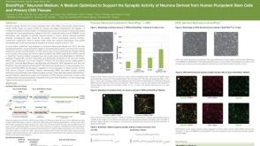
BrainPhys™神经元培养基
BrainPhys™神经元培养基和SM1试剂盒
BrainPhys™ 神经元培养基N2-A和SM1试剂盒
BrainPhys™原代神经元试剂盒
BrainPhys™ hPSC 神经元试剂盒
NeuroCult™ SM1 神经添加物
Kayama T et al. (JAN 2018)
Biochemical and Biophysical Research Communications 495 1 1028--1033
Temporally coordinated spiking activity of human induced pluripotent stem cell-derived neurons co-cultured with astrocytes
In culture conditions,human induced-pluripotent stem cells (hiPSC)-derived neurons form synaptic connections with other cells and establish neuronal networks,which are expected to be an in vitro model system for drug discovery screening and toxicity testing. While early studies demonstrated effects of co-culture of hiPSC-derived neurons with astroglial cells on survival and maturation of hiPSC-derived neurons,the population spiking patterns of such hiPSC-derived neurons have not been fully characterized. In this study,we analyzed temporal spiking patterns of hiPSC-derived neurons recorded by a multi-electrode array system. We discovered that specific sets of hiPSC-derived neurons co-cultured with astrocytes showed more frequent and highly coherent non-random synchronized spike trains and more dynamic changes in overall spike patterns over time. These temporally coordinated spiking patterns are physiological signs of organized circuits of hiPSC-derived neurons and suggest benefits of co-culture of hiPSC-derived neurons with astrocytes.
View Publication

 EasySep™小鼠TIL(CD45)正选试剂盒


 沪公网安备31010102008431号