Lin H et al. (JAN 2017)
Neuro-oncology 19 1 43--54
Fatty acid oxidation is required for the respiration and proliferation of malignant glioma cells.
BACKGROUND Glioma is the most common form of primary malignant brain tumor in adults,with approximately 4 cases per 100 000 people each year. Gliomas,like many tumors,are thought to primarily metabolize glucose for energy production; however,the reliance upon glycolysis has recently been called into question. In this study,we aimed to identify the metabolic fuel requirements of human glioma cells. METHODS We used database searches and tissue culture resources to evaluate genotype and protein expression,tracked oxygen consumption rates to study metabolic responses to various substrates,performed histochemical techniques and fluorescence-activated cell sorting-based mitotic profiling to study cellular proliferation rates,and employed an animal model of malignant glioma to evaluate a new therapeutic intervention. RESULTS We observed the presence of enzymes required for fatty acid oxidation within human glioma tissues. In addition,we demonstrated that this metabolic pathway is a major contributor to aerobic respiration in primary-cultured cells isolated from human glioma and grown under serum-free conditions. Moreover,inhibiting fatty acid oxidation reduces proliferative activity in these primary-cultured cells and prolongs survival in a syngeneic mouse model of malignant glioma. CONCLUSIONS Fatty acid oxidation enzymes are present and active within glioma tissues. Targeting this metabolic pathway reduces energy production and cellular proliferation in glioma cells. The drug etomoxir may provide therapeutic benefit to patients with malignant glioma. In addition,the expression of fatty acid oxidation enzymes may provide prognostic indicators for clinical practice.
View Publication
产品号#:
05750
05751
产品名:
NeuroCult™ NS-A 基础培养基(人)
NeuroCult™ NS-A 扩增试剂盒(人)
Li Y et al. (JAN 2016)
Journal of virology 90 7 3385--99
Ecotropic Murine Leukemia Virus Infection of Glial Progenitors Interferes with Oligodendrocyte Differentiation: Implications for Neurovirulence.
UNLABELLED Certain murine leukemia viruses (MLVs) are capable of inducing fatal progressive spongiform motor neuron disease in mice that is largely mediated by viral Env glycoprotein expression within central nervous system (CNS) glia. While the etiologic mechanisms and the glial subtypes involved remain unresolved,infection of NG2 glia was recently observed to correlate spatially and temporally with altered neuronal physiology and spongiogenesis. Since one role of NG2 cells is to serve as oligodendrocyte (OL) progenitor cells (OPCs),we examined here whether their infection by neurovirulent (FrCasE) or nonneurovirulent (Fr57E) ecotropic MLVs influenced their viability and/or differentiation. Here,we demonstrate that OPCs,but not OLs,are major CNS targets of both FrCasE and Fr57E. We also show that MLV infection of neural progenitor cells (NPCs) in culture did not affect survival,proliferation,or OPC progenitor marker expression but suppressed certain glial differentiation markers. Assessment of glial differentiation in vivo using transplanted transgenic NPCs showed that,while MLVs did not affect cellular engraftment or survival,they did inhibit OL differentiation,irrespective of MLV neurovirulence. In addition,in chimeric brains,where FrCasE-infected NPC transplants caused neurodegeneration,the transplanted NPCs proliferated. These results suggest that MLV infection is not directly cytotoxic to OPCs but rather acts to interfere with OL differentiation. Since both FrCasE and Fr57E viruses restrict OL differentiation but only FrCasE induces overt neurodegeneration,restriction of OL maturation alone cannot account for neuropathogenesis. Instead neurodegeneration may involve a two-hit scenario where interference with OPC differentiation combined with glial Env-induced neuronal hyperexcitability precipitates disease. IMPORTANCE A variety of human and animal retroviruses are capable of causing central nervous system (CNS) neurodegeneration manifested as motor and cognitive deficits. These retroviruses infect a variety of CNS cell types; however,the specific role each cell type plays in neuropathogenesis remains to be established. The NG2 glia,whose CNS functions are only now emerging,are a newly appreciated viral target in murine leukemia virus (MLV)-induced neurodegeneration. Since one role of NG2 glia is that of oligodendrocyte progenitor cells (OPCs),we investigated here whether their infection by the neurovirulent MLV FrCasE contributed to neurodegeneration by affecting OPC viability and/or development. Our results show that both neurovirulent and nonneurovirulent MLVs interfere with oligodendrocyte differentiation. Thus,NG2 glial infection could contribute to neurodegeneration by preventing myelin formation and/or repair and by suspending OPCs in a state of persistent susceptibility to excitotoxic insult mediated by neurovirulent virus effects on other glial subtypes.
View Publication
Kayama T et al. (JAN 2018)
Biochemical and Biophysical Research Communications 495 1 1028--1033
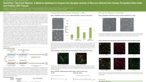
Temporally coordinated spiking activity of human induced pluripotent stem cell-derived neurons co-cultured with astrocytes
In culture conditions,human induced-pluripotent stem cells (hiPSC)-derived neurons form synaptic connections with other cells and establish neuronal networks,which are expected to be an in vitro model system for drug discovery screening and toxicity testing. While early studies demonstrated effects of co-culture of hiPSC-derived neurons with astroglial cells on survival and maturation of hiPSC-derived neurons,the population spiking patterns of such hiPSC-derived neurons have not been fully characterized. In this study,we analyzed temporal spiking patterns of hiPSC-derived neurons recorded by a multi-electrode array system. We discovered that specific sets of hiPSC-derived neurons co-cultured with astrocytes showed more frequent and highly coherent non-random synchronized spike trains and more dynamic changes in overall spike patterns over time. These temporally coordinated spiking patterns are physiological signs of organized circuits of hiPSC-derived neurons and suggest benefits of co-culture of hiPSC-derived neurons with astrocytes.
View Publication
产品号#:
05790
05792
05793
05794
05795
产品名:
BrainPhys™神经元培养基
BrainPhys™神经元培养基和SM1试剂盒
BrainPhys™ 神经元培养基N2-A和SM1试剂盒
BrainPhys™原代神经元试剂盒
BrainPhys™ hPSC 神经元试剂盒
Lawn S et al. (FEB 2015)
The Journal of biological chemistry 290 6 3814--24
Neurotrophin signaling via TrkB and TrkC receptors promotes the growth of brain tumor-initiating cells.
Neurotrophins and their receptors are frequently expressed in malignant gliomas,yet their functions are largely unknown. Previously,we have shown that p75 neurotrophin receptor is required for glioma invasion and proliferation. However,the role of Trk receptors has not been examined. In this study,we investigated the importance of TrkB and TrkC in survival of brain tumor-initiating cells (BTICs). Here,we show that human malignant glioma tissues and also tumor-initiating cells isolated from fresh human malignant gliomas express the neurotrophin receptors TrkB and TrkC,not TrkA,and they also express neurotrophins NGF,BDNF,and neurotrophin 3 (NT3). Specific activation of TrkB and TrkC receptors by ligands BDNF and NT3 enhances tumor-initiating cell viability through activation of ERK and Akt pathways. Conversely,TrkB and TrkC knockdown or pharmacologic inhibition of Trk signaling decreases neurotrophin-dependent ERK activation and BTIC growth. Further,pharmacological inhibition of both ERK and Akt pathways blocked BDNF,and NT3 stimulated BTIC survival. Importantly,attenuation of BTIC growth by EGFR inhibitors could be overcome by activation of neurotrophin signaling,and neurotrophin signaling is sufficient for long term BTIC growth as spheres in the absence of EGF and FGF. Our results highlight a novel role for neurotrophin signaling in brain tumor and suggest that Trks could be a target for combinatorial treatment of malignant glioma.
View Publication
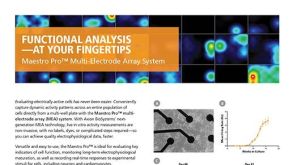
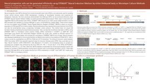
On-demand optogenetic activation of human stem-cell-derived neurons
The widespread application of human stem-cell-derived neurons for functional studies is impeded by complicated differentiation protocols,immaturity,and deficient optogene expression as stem cells frequently lose transgene expression over time. Here we report a simple but precise Cre-loxP-based strategy for generating conditional,and thereby stable,optogenetic human stem-cell lines. These cells can be easily and efficiently differentiated into functional neurons,and optogene expression can be triggered by administering Cre protein to the cultures. This conditional expression system may be applied to stem-cell-derived neurons whenever timed transgene expression could help to overcome silencing at the stem-cell level.
View Publication

 EasySep™小鼠TIL(CD45)正选试剂盒


 沪公网安备31010102008431号