
 EasySep™小鼠TIL(CD45)正选试剂盒


技术资料
-
 产品手册Highway1â„¢: Fast, Gentle, and Automated Cell Sorting for Every Lab
品牌:
Highway1
发布日期: 04/14/2026 -
 科学海报Mechanoporation-Based Intracellular Delivery of Functional Molecules for Primary Immune Cell Engineering
Conference:
AAI 2026
-
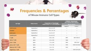 挂图Frequencies and Percentages of Mouse Immune Cell Types List of the frequencies of over 25 immune cell types in C57BL/6 mice


 沪公网安备31010102008431号