
 EasySep™小鼠TIL(CD45)正选试剂盒


技术资料
-
 产品手册Highway1™: Fast, Gentle, and Automated Cell Sorting for Every Lab
品牌:
Highway1
发布日期: 04/14/2026 -
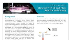 技术公告Protocol for Semi-Solid Hybridoma Cloning Using ClonaCell™-HY Medium in a 96-Well Plate
细胞类型:
杂交瘤细胞
发布日期: 05/30/2023 -
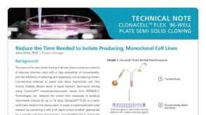 技术公告Protocol for Producing Monoclonal Cell Lines Using ClonaCell™ FLEX Semi-Solid Medium
细胞类型:
CHO细胞,杂交瘤细胞
发布日期: 05/01/2023 -
-


 沪公网安备31010102008431号