Zhu TS et al. (SEP 2011)
Cancer research 71 18 6061--72
Endothelial cells create a stem cell niche in glioblastoma by providing NOTCH ligands that nurture self-renewal of cancer stem-like cells.
One important function of endothelial cells in glioblastoma multiforme (GBM) is to create a niche that helps promote self-renewal of cancer stem-like cells (CSLC). However,the underlying molecular mechanism for this endothelial function is not known. Since activation of NOTCH signaling has been found to be required for propagation of GBM CSLCs,we hypothesized that the GBM endothelium may provide the source of NOTCH ligands. Here,we report a corroboration of this concept with a demonstration that NOTCH ligands are expressed in endothelial cells adjacent to NESTIN and NOTCH receptor-positive cancer cells in primary GBMs. Coculturing human brain microvascular endothelial cells (hBMEC) or NOTCH ligand with GBM neurospheres promoted GBM cell growth and increased CSLC self-renewal. Notably,RNAi-mediated knockdown of NOTCH ligands in hBMECs abrogated their ability to induce CSLC self-renewal and GBM tumor growth,both in vitro and in vivo. Thus,our findings establish that NOTCH activation in GBM CSLCs is driven by juxtacrine signaling between tumor cells and their surrounding endothelial cells in the tumor microenvironment,suggesting that targeting both CSLCs and their niche may provide a novel strategy to deplete CSLCs and improve GBM treatment.
View Publication
产品号#:
05750
05751
05752
产品名:
NeuroCult™ NS-A 基础培养基(人)
NeuroCult™ NS-A 扩增试剂盒(人)
NeuroCult™ NS-A 分化试剂盒(人)
Binder ZA et al. ( 2013)
PloS one 8 10 e75945
Podocalyxin-like protein is expressed in glioblastoma multiforme stem-like cells and is associated with poor outcome.
Glioblastoma multiforme (GBM) is the most common primary malignant adult brain tumor and is associated with poor survival. Recently,stem-like cell populations have been identified in numerous malignancies including GBM. To identify genes whose expression is changed with differentiation,we compared transcript profiles from a GBM oncosphere line before and after differentiation. Bioinformatic analysis of the gene expression profiles identified podocalyxin-like protein (PODXL),a protein highly expressed in human embryonic stem cells,as a potential marker of undifferentiated GBM stem-like cells. The loss of PODXL expression upon differentiation of GBM stem-like cell lines was confirmed by quantitative real-time PCR and flow cytometry. Analytical flow cytometry of numerous GBM oncosphere lines demonstrated PODXL expression in all lines examined. Knockdown studies and flow cytometric cell sorting experiments demonstrated that PODXL is involved in GBM stem-like cell proliferation and oncosphere formation. Compared to PODXL-negative cells,PODXL-positive cells had increased expression of the progenitor/stem cell markers Musashi1,SOX2,and BMI1. Finally,PODXL expression directly correlated with increasing glioma grade and was a marker for poor outcome in patients with GBM. In summary,we have demonstrated that PODXL is expressed in GBM stem-like cells and is involved in cell proliferation and oncosphere formation. Moreover,high PODXL expression correlates with increasing glioma grade and decreased overall survival in patients with GBM.
View Publication
产品号#:
05750
05751
产品名:
NeuroCult™ NS-A 基础培养基(人)
NeuroCult™ NS-A 扩增试剂盒(人)
Chung D et al. (JAN 2014)
The Veterinary Journal 199 1 123--130
Effect of hypoxia on generation of neurospheres from adipose tissue-derived canine mesenchymal stromal cells
Adipose tissue-derived mesenchymal stromal cells (AT-MSCs) are good candidates for cell therapy due to the accessibility of fat tissue and the abundance of AT-MSCs therein. Neurospheres are free-floating spherical condensations of cells with neural stem/progenitor cell (NSPC) characteristics that can be derived from AT-MSCs. The aims of this study were to examine the influence of oxygen (O2) tension on generation of neurospheres from canine AT-MSCs (AT-cMSCs) and to develop a hypoxic cell culture system to enhance the survival and therapeutic benefit of generated neurospheres. AT-cMSCs were cultured under varying oxygen tensions (1%,5% and 21%) in a neurosphere culture system. Neurosphere number and area were evaluated and NSPC markers were quantified using real-time quantitative PCR (qPCR). Effects of oxygen on neurosphere expression of hypoxia inducible factor 1,α subunit (HIF1A) and its target genes,erythropoietin receptor (EPOR),chemokine (C-X-C motif) receptor 4 (CXCR4) and vascular endothelial growth factor (VEGF),were quantified by qPCR. Neural differentiation potential was evaluated in 21% O2 by cell morphology and qPCR. Neurospheres were successfully generated from AT-cMSCs at all O2 tensions. Expression of nestin mRNA (NES) was significantly increased after neurosphere culture and was significantly higher in 1% O2 compared to 5% and 21% O2. Neurospheres cultured in 1% O2 had significantly increased levels of VEGF and EPOR. There was a significant increase in CXCR4 expression in neurospheres generated at all O2 tensions. Neurosphere culture under hypoxia had no negative effect on subsequent neural differentiation. This study suggests that generation of neurospheres under hypoxia could be beneficial when considering these cells for neurological cell therapies.
View Publication
产品号#:
05750
05751
05752
产品名:
NeuroCult™ NS-A 基础培养基(人)
NeuroCult™ NS-A 扩增试剂盒(人)
NeuroCult™ NS-A 分化试剂盒(人)
Halvorson KG et al. ( 2015)
PloS one 10 3 e0118926
A high-throughput in vitro drug screen in a genetically engineered mouse model of diffuse intrinsic pontine glioma identifies BMS-754807 as a promising therapeutic agent.
Diffuse intrinsic pontine gliomas (DIPGs) represent a particularly lethal type of pediatric brain cancer with no effective therapeutic options. Our laboratory has previously reported the development of genetically engineered DIPG mouse models using the RCAS/tv-a system,including a model driven by PDGF-B,H3.3K27M,and p53 loss. These models can serve as a platform in which to test novel therapeutics prior to the initiation of human clinical trials. In this study,an in vitro high-throughput drug screen as part of the DIPG preclinical consortium using cell-lines derived from our DIPG models identified BMS-754807 as a drug of interest in DIPG. BMS-754807 is a potent and reversible small molecule multi-kinase inhibitor with many targets including IGF-1R,IR,MET,TRKA,TRKB,AURKA,AURKB. In vitro evaluation showed significant cytotoxic effects with an IC50 of 0.13 μM,significant inhibition of proliferation at a concentration of 1.5 μM,as well as inhibition of AKT activation. Interestingly,IGF-1R signaling was absent in serum-free cultures from the PDGF-B; H3.3K27M; p53 deficient model suggesting that the antitumor activity of BMS-754807 in this model is independent of IGF-1R. In vivo,systemic administration of BMS-754807 to DIPG-bearing mice did not prolong survival. Pharmacokinetic analysis demonstrated that tumor tissue drug concentrations of BMS-754807 were well below the identified IC50,suggesting that inadequate drug delivery may limit in vivo efficacy. In summary,an unbiased in vitro drug screen identified BMS-754807 as a potential therapeutic agent in DIPG,but BMS-754807 treatment in vivo by systemic delivery did not significantly prolong survival of DIPG-bearing mice.
View Publication
产品号#:
05700
产品名:
NeuroCult™ 基础培养基(小鼠和大鼠)
Duan S et al. (DEC 2015)
Nature communications 6 10068
PTEN deficiency reprogrammes human neural stem cells towards a glioblastoma stem cell-like phenotype.
PTEN is a tumour suppressor frequently mutated in many types of cancers. Here we show that targeted disruption of PTEN leads to neoplastic transformation of human neural stem cells (NSCs),but not mesenchymal stem cells. PTEN-deficient NSCs display neoplasm-associated metabolic and gene expression profiles and generate intracranial tumours in immunodeficient mice. PTEN is localized to the nucleus in NSCs,binds to the PAX7 promoter through association with cAMP responsive element binding protein 1 (CREB)/CREB binding protein (CBP) and inhibits PAX7 transcription. PTEN deficiency leads to the upregulation of PAX7,which in turn promotes oncogenic transformation of NSCs and instates 'aggressiveness' in human glioblastoma stem cells. In a large clinical database,we find increased PAX7 levels in PTEN-deficient glioblastoma. Furthermore,we identify that mitomycin C selectively triggers apoptosis in NSCs with PTEN deficiency. Together,we uncover a potential mechanism of how PTEN safeguards NSCs,and establish a cellular platform to identify factors involved in NSC transformation,potentially permitting personalized treatment of glioblastoma.
View Publication
产品号#:
05700
05701
05702
05750
05850
05857
05870
05875
85850
85857
85870
85875
产品名:
NeuroCult™ 基础培养基(小鼠和大鼠)
NeuroCult™ 扩增添加物(小鼠和大鼠)
NeuroCult™扩增试剂盒(小鼠和大鼠)
NeuroCult™ NS-A 基础培养基(人)
mTeSR™1
mTeSR™1
Setty M et al. (JAN 2012)
Molecular systems biology 8 605
Inferring transcriptional and microRNA-mediated regulatory programs in glioblastoma.
Large-scale cancer genomics projects are profiling hundreds of tumors at multiple molecular layers,including copy number,mRNA and miRNA expression,but the mechanistic relationships between these layers are often excluded from computational models. We developed a supervised learning framework for integrating molecular profiles with regulatory sequence information to reveal regulatory programs in cancer,including miRNA-mediated regulation. We applied our approach to 320 glioblastoma profiles and identified key miRNAs and transcription factors as common or subtype-specific drivers of expression changes. We confirmed that predicted gene expression signatures for proneural subtype regulators were consistent with in vivo expression changes in a PDGF-driven mouse model. We tested two predicted proneural drivers,miR-124 and miR-132,both underexpressed in proneural tumors,by overexpression in neurospheres and observed a partial reversal of corresponding tumor expression changes. Computationally dissecting the role of miRNAs in cancer may ultimately lead to small RNA therapeutics tailored to subtype or individual.
View Publication
产品号#:
05750
05751
产品名:
NeuroCult™ NS-A 基础培养基(人)
NeuroCult™ NS-A 扩增试剂盒(人)
Buczkowicz P et al. (MAY 2013)
Brain pathology (Zurich,Switzerland) 23 3 244--53
Aurora kinase B is a potential therapeutic target in pediatric diffuse intrinsic pontine glioma.
Pediatric high-grade astrocytomas (HGAs) account for 15-20% of all pediatric central nervous system tumors. These neoplasms predominantly involve the supratentorial hemispheres or the pons--diffuse intrinsic pontine gliomas (DIPG). Assumptions that pediatric HGAs are biologically similar to adult HGAs have recently been challenged,and the development of effective therapeutic modalities for DIPG and supratentorial HGA hinges on a better understanding of their biological properties. Here,20 pediatric HGAs (9 DIPGs and 11 supratentorial HGAs) were subject to gene expression profiling following approval by the research ethics board at our institution. Many of these tumors showed expression signatures composed of genes that promote G1/S and G2/M cell cycle progression. In particular,Aurora kinase B (AURKB) was consistently and highly overexpressed in 6/9 DIPGs and 8/11 HGAs. Array data were validated using quantitative real-time PCR and immunohistochemistry,as well as cross-validation of our data set with previously published series. Inhibition of Aurora B activity in DIPG and in pediatric HGA cell lines resulted in growth arrest accompanied by morphological changes,cell cycle aberrations,nuclear fractionation and polyploidy as well as a reduction in colony formation. Our data highlight Aurora B as a potential therapeutic target in DIPG.
View Publication
Gundemir S et al. (SEP 2016)
Neuro-Oncology now157
The complex role of transglutaminase 2 in glioblastoma proliferation
BACKGROUND Glioblastomas (GBMs) are a heterogeneous group of primary brain tumors. These tumors are resistant to therapeutic interventions and invariably recur after surgical resection. The multifunctional protein transglutaminase 2 (TG2) has been shown to promote cell survival in a number of different tumors. There is also evidence that TG2 may be a pro-survival factor in GBMs. However,the roles that TG2 plays in facilitating GBM survival and proliferation have not yet been clearly delineated . METHODS The functions of TG2 are often cell- and context-specific. Therefore,in this study we examined the ability of TG2 to facilitate GBM proliferation using colony formation assays and 5-ethynyl-2'-deoxyuridine (EdU) incorporation in several different GBM cell lines as well as neurospheres derived from patient tumors representing the 3 major subtypes of GBM tumors (mesenchymal,proneural,and classical) and maintained in the absence of serum. TG2 knockdown or selective TG2 inhibitors were used to modulate TG2 expression and activity. RESULTS We show that TG2 plays differential roles in the proliferative process depending on the cell type. In most,but not all,GBM models TG2 plays a crucial role in the proliferative process,and some but not all TG2 inhibitors were highly effective at reducing proliferation in a large subset of the GBM models. CONCLUSION Our results show that TG2 plays an important-but notoriously context-specific-role in GBM cell biology. Nonetheless,as future studies unravel the genetic fingerprints" that make TG2 inhibitors effective this information could be exploited to develop TG2 inhibitors into personalized GBM therapies.
View Publication
产品号#:
05750
05751
产品名:
NeuroCult™ NS-A 基础培养基(人)
NeuroCult™ NS-A 扩增试剂盒(人)
Marigil M et al. (JAN 2017)
PloS one 12 1 e0170501
Development of a DIPG Orthotopic Model in Mice Using an Implantable Guide-Screw System.
OBJECTIVE In this work we set to develop and to validate a new in vivo frameless orthotopic Diffuse Intrinsic Pontine Glioma (DIPG) model based in the implantation of a guide-screw system. METHODS It consisted of a guide-screw also called bolt,a Hamilton syringe with a 26-gauge needle and an insulin-like 15-gauge needle. The guide screw is 2.6 mm in length and harbors a 0.5 mm central hole which accepts the needle of the Hamilton syringe avoiding a theoretical displacement during insertion. The guide-screw is fixed on the mouse skull according to the coordinates: 1mm right to and 0.8 mm posterior to lambda. To reach the pons the Hamilton syringe is adjusted to a 6.5 mm depth using a cuff that serves as a stopper. This system allows delivering not only cells but also any kind of intratumoral chemotherapy,antibodies or gene/viral therapies. RESULTS The guide-screw was successfully implanted in 10 immunodeficient mice and the animals were inoculated with DIPG human cell lines during the same anesthetic period. All the mice developed severe neurologic symptoms and had a median overall survival of 95 days ranging the time of death from 81 to 116 days. Histopathological analysis confirmed tumor into the pons in all animals confirming the validity of this model. CONCLUSION Here we presented a reproducible and frameless DIPG model that allows for rapid evaluation of tumorigenicity and efficacy of chemotherapeutic or gene therapy products delivered intratumorally to the pons.
View Publication

 EasySep™小鼠TIL(CD45)正选试剂盒


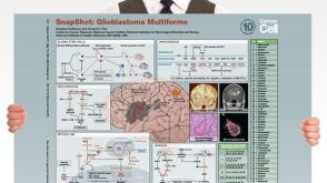 挂图SnapShot: Glioblastoma Multiforme Overview of the key concepts and mechanisms in glioblastoma multiforme biology
 沪公网安备31010102008431号