Takayama Y and Kida YS (FEB 2016)
PloS one 11 2 e0148559
In Vitro Reconstruction of Neuronal Networks Derived from Human iPS Cells Using Microfabricated Devices.
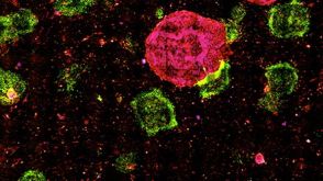
Morphology and function of the nervous system is maintained via well-coordinated processes both in central and peripheral nervous tissues,which govern the homeostasis of organs/tissues. Impairments of the nervous system induce neuronal disorders such as peripheral neuropathy or cardiac arrhythmia. Although further investigation is warranted to reveal the molecular mechanisms of progression in such diseases,appropriate model systems mimicking the patient-specific communication between neurons and organs are not established yet. In this study,we reconstructed the neuronal network in vitro either between neurons of the human induced pluripotent stem (iPS) cell derived peripheral nervous system (PNS) and central nervous system (CNS),or between PNS neurons and cardiac cells in a morphologically and functionally compartmentalized manner. Networks were constructed in photolithographically microfabricated devices with two culture compartments connected by 20 microtunnels. We confirmed that PNS and CNS neurons connected via synapses and formed a network. Additionally,calcium-imaging experiments showed that the bundles originating from the PNS neurons were functionally active and responded reproducibly to external stimuli. Next,we confirmed that CNS neurons showed an increase in calcium activity during electrical stimulation of networked bundles from PNS neurons in order to demonstrate the formation of functional cell-cell interactions. We also confirmed the formation of synapses between PNS neurons and mature cardiac cells. These results indicate that compartmentalized culture devices are promising tools for reconstructing network-wide connections between PNS neurons and various organs,and might help to understand patient-specific molecular and functional mechanisms under normal and pathological conditions.
View Publication
产品号#:
05850
05857
05870
05875
85850
85857
85870
85875
产品名:
mTeSR™1
mTeSR™1
Yan Y et al. (FEB 2015)
1341 257--284
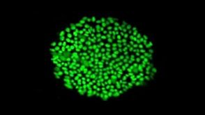
Generation of Neural Progenitor Spheres from Human Pluripotent Stem Cells in a Suspension Bioreactor
Conventional two-dimensional (2-D) culture systems cannot provide large numbers of human pluripotent stem cells (hPSCs) and their derivatives that are demanded for commercial and clinical applications in in vitro drug screening,disease modeling,and potentially cell therapy. The technologies that support three-dimensional (3-D) suspension culture,such as a stirred bioreactor,are generally considered as promising approaches to produce the required cells. Recently,suspension bioreactors have also been used to generate mini-brain-like structure from hPSCs for disease modeling,showing the important role of bioreactor in stem cell culture. This chapter describes a detailed culture protocol for neural commitment of hPSCs into neural progenitor cell (NPC) spheres using a spinner bioreactor. The basic steps to prepare hPSCs for bioreactor inoculation are illustrated from cell thawing to cell propagation. The method for generating NPCs from hPSCs in the spinner bioreactor along with the static control is then described. The protocol in this study can be applied to the generation of NPCs from hPSCs for further neural subtype specification,3-D neural tissue development,or potential preclinical studies or clinical applications in neurological diseases.
View Publication
产品号#:
05850
05857
05870
05875
72302
72304
72307
72308
85850
85857
85870
85875
100-1044
产品名:
Y-27632(二盐酸盐)
Y-27632(二盐酸盐)
Y-27632(二盐酸盐)
Y-27632(二盐酸盐)
mTeSR™1
mTeSR™1
Y-27632(二盐酸盐)
Wang R et al. (DEC 2015)
BMC cancer 16 1 56
Fusion with stem cell makes the hepatocellular carcinoma cells similar to liver tumor-initiating cells.
BACKGROUND Cell fusion is a fast and highly efficient technique for cells to acquire new properties. The fusion of somatic cells with stem cells can reprogram somatic cells to a pluripotent state. Our research on the fusion of stem cells and cancer cells demonstrates that the fused cells can exhibit stemness and cancer cell-like characteristics. Thus,tumor-initiating cell-like cells are generated. METHODS We employed laser-induced single-cell fusion technique to fuse the hepatocellular carcinoma cells and human embryonic stem cells (hESC). Real-time RT-PCR,flow cytometry and in vivo tumorigenicity assay were adopted to identify the gene expression difference. RESULTS We successfully produced a fused cell line that coalesces the gene expression information of hepatocellular carcinoma cells and stem cells. Experimental results showed that the fused cells expressed cancer and stemness markers as well as exhibited increased resistance to drug treatment and enhanced tumorigenesis. CONCLUSIONS Fusion with stem cells transforms liver cancer cells into tumor initiating-like cells. Results indicate that fusion between cancer cell and stem cell may generate tumor initiating-like cells.
View Publication
产品号#:
05850
05857
05870
05875
85850
85857
85870
85875
产品名:
mTeSR™1
mTeSR™1
Seibler P et al. (APR 2011)
The Journal of neuroscience : the official journal of the Society for Neuroscience 31 16 5970--6
Mitochondrial Parkin recruitment is impaired in neurons derived from mutant PINK1 induced pluripotent stem cells.
Genetic Parkinson disease (PD) has been associated with mutations in PINK1,a gene encoding a mitochondrial kinase implicated in the regulation of mitochondrial degradation. While the studies so far examined PINK1 function in non-neuronal systems or through PINK1 knockdown approaches,there is an imperative to examine the role of endogenous PINK1 in appropriate human-derived and biologically relevant cell models. Here we report the generation of induced pluripotent stem (iPS) cells from skin fibroblasts taken from three PD patients with nonsense (c.1366CtextgreaterT; p.Q456X) or missense (c.509TtextgreaterG; p.V170G) mutations in the PINK1 gene. These cells were differentiated into dopaminergic neurons that upon mitochondrial depolarization showed impaired recruitment of lentivirally expressed Parkin to mitochondria,increased mitochondrial copy number,and upregulation of PGC-1α,an important regulator of mitochondrial biogenesis. Importantly,these alterations were corrected by lentiviral expression of wild-type PINK1 in mutant iPS cell-derived PINK1 neurons. In conclusion,our studies suggest that fibroblasts from genetic PD can be reprogrammed and differentiated into neurons. These neurons exhibit distinct phenotypes that should be amenable to further mechanistic studies in this relevant biological context.
View Publication
产品号#:
05850
05857
05870
05875
36254
85850
85857
85870
85875
产品名:
DMEM/F-12 with 15 mM HEPES
mTeSR™1
mTeSR™1
Moore RN et al. (JAN 2012)
Stem cells and development 21 1 30--41
E-cadherin-expressing feeder cells promote neural lineage restriction of human embryonic stem cells.
Human embryonic stem cells (hESCs) represent a promising source of tissues of different cell lineages because of their high degree of self-renewal and their unique ability to give rise to most somatic cell lineages. In this article,we report on a new approach to differentiate hESCs into neural stem cells that can be differentiated further into neuronal restricted cells. We have rapidly and efficiently differentiated hESCs into neural stem cells by presenting the cell adhesion molecule,E-cadherin,to undifferentiated hESCs via E-cadherin transfected fibroblast monolayers. The neural restricted progenitor cells rapidly express nestin and beta-III-tubulin,but not glial fibrillary acidic protein (GFAP) during the 1-week E-cadherin induction phase,suggesting that E-cadherin promotes rapid neuronal differentiation. Further,these cells are able to achieve enhanced neuronal differentiation with the addition of exogenous growth factors. Cadherin-induced hESCs show a loss in Oct4 and nestin expression associated with positive staining for vimentin,neurofilament,and neural cell adhesion molecule. Moreover,blocking by functional E-cadherin antibody and failure of paracrine stimulation suggested that direct E-cadherin engagement is necessary to induce neural restriction. By providing hESCs with molecular cues to promote differentiation,we are able to utilize a specific cell-cell adhesion molecule,E-cadherin,to influence the nature and degree of neural specialization.
View Publication
产品号#:
05850
05857
05870
05875
85850
85857
85870
85875
产品名:
mTeSR™1
mTeSR™1
Xie L et al. (APR 2011)
The EMBO journal 30 8 1473--84
Although regulation of histone methylation is believed to contribute to embryonic stem cell (ESC) self-renewal,the mechanisms remain obscure. We show here that the histone H3 trimethyl lysine 4 (H3K4me3) demethylase,KDM5B,is a downstream Nanog target and critical for ESC self-renewal. Although KDM5B is believed to function as a promoter-bound repressor,we find that it paradoxically functions as an activator of a gene network associated with self-renewal. ChIP-Seq reveals that KDM5B is predominantly targeted to intragenic regions and that it is recruited to H3K36me3 via an interaction with the chromodomain protein MRG15. Depletion of KDM5B or MRG15 increases intragenic H3K4me3,increases cryptic intragenic transcription,and inhibits transcriptional elongation of KDM5B target genes. We propose that KDM5B activates self-renewal-associated gene expression by repressing cryptic initiation and maintaining an H3K4me3 gradient important for productive transcriptional elongation.
View Publication
产品号#:
05850
05857
05870
05875
85850
85857
85870
85875
产品名:
mTeSR™1
mTeSR™1
Zhang J et al. (NOV 2011)
Stem Cell Reviews and Reports 7 4 987--996
Electrically Guiding Migration of Human Induced Pluripotent Stem Cells
A major road-block in stem cell therapy is the poor homing and integration of transplanted stem cells with the targeted host tissue. Human induced pluripotent stem (hiPS) cells are considered an excellent alternative to embryonic stem (ES) cells and we tested the feasibility of using small,physiological electric fields (EFs) to guide hiPS cells to their target. Applied EFs stimulated and guided migration of cultured hiPS cells toward the anode,with a stimulation threshold of textless30 mV/mm; in three-dimensional (3D) culture hiPS cells remained stationary,whereas in an applied EF they migrated directionally. This is of significance as the therapeutic use of hiPS cells occurs in a 3D environment. EF exposure did not alter expression of the pluripotency markers SSEA-4 and Oct-4 in hiPS cells. We compared EF-directed migration (galvanotaxis) of hiPS cells and hES cells and found that hiPS cells showed greater sensitivity and directedness than those of hES cells in an EF,while hES cells migrated toward cathode. Rho-kinase (ROCK) inhibition,a method to aid expansion and survival of stem cells,significantly increased the motility,but reduced directionality of iPS cells in an EF by 70-80%. Thus,our study has revealed that physiological EF is an effective guidance cue for the migration of hiPS cells in either 2D or 3D environments and that will occur in a ROCK-dependent manner. Our current finding may lead to techniques for applying EFs in vivo to guide migration of transplanted stem cells.
View Publication
产品号#:
05850
05857
05870
05875
85850
85857
85870
85875
产品名:
mTeSR™1
mTeSR™1
Mortensen M et al. (MAR 2011)
The Journal of experimental medicine 208 3 455--67
The autophagy protein Atg7 is essential for hematopoietic stem cell maintenance.
The role of autophagy,a lysosomal degradation pathway which prevents cellular damage,in the maintenance of adult mouse hematopoietic stem cells (HSCs) remains unknown. Although normal HSCs sustain life-long hematopoiesis,malignant transformation of HSCs leads to leukemia. Therefore,mechanisms protecting HSCs from cellular damage are essential to prevent hematopoietic malignancies. In this study,we crippled autophagy in HSCs by conditionally deleting the essential autophagy gene Atg7 in the hematopoietic system. This resulted in the loss of normal HSC functions,a severe myeloproliferation,and death of the mice within weeks. The hematopoietic stem and progenitor cell compartment displayed an accumulation of mitochondria and reactive oxygen species,as well as increased proliferation and DNA damage. HSCs within the Lin(-)Sca-1(+)c-Kit(+) (LSK) compartment were significantly reduced. Although the overall LSK compartment was expanded,Atg7-deficient LSK cells failed to reconstitute the hematopoietic system of lethally irradiated mice. Consistent with loss of HSC functions,the production of both lymphoid and myeloid progenitors was impaired in the absence of Atg7. Collectively,these data show that Atg7 is an essential regulator of adult HSC maintenance.
View Publication
产品号#:
03434
03444
产品名:
MethoCult™ GF M3434
MethoCult™ GF M3434
Goossens S et al. (MAY 2011)
Blood 117 21 5620--30
The EMT regulator Zeb2/Sip1 is essential for murine embryonic hematopoietic stem/progenitor cell differentiation and mobilization.
Zeb2 (Sip1/Zfhx1b) is a member of the zinc-finger E-box-binding (ZEB) family of transcriptional repressors previously demonstrated to regulate epithelial-to-mesenchymal transition (EMT) processes during embryogenesis and tumor progression. We found high Zeb2 mRNA expression levels in HSCs and hematopoietic progenitor cells (HPCs),and examined Zeb2 function in hematopoiesis through a conditional deletion approach using the Tie2-Cre and Vav-iCre recombination mouse lines. Detailed cellular analysis demonstrated that Zeb2 is dispensable for hematopoietic cluster and HSC formation in the aorta-gonadomesonephros region of the embryo,but is essential for normal HSC/HPC differentiation. In addition,Zeb2-deficient HSCs/HPCs fail to properly colonize the fetal liver and/or bone marrow and show enhanced adhesive properties associated with increased β1 integrin and Cxcr4 expression. Moreover,deletion of Zeb2 resulted in embryonic (Tie2-Cre) and perinatal (Vav-icre) lethality due to severe cephalic hemorrhaging and decreased levels of angiopoietin-1 and,subsequently,improper pericyte coverage of the cephalic vasculature. These results reveal essential roles for Zeb2 in embryonic hematopoiesis and are suggestive of a role for Zeb2 in hematopoietic-related pathologies in the adult.
View Publication

 EasySep™小鼠TIL(CD45)正选试剂盒


 沪公网安备31010102008431号