Hartfield EM et al. (FEB 2014)
PLoS ONE 9 2 e87388
Physiological characterisation of human iPS-derived dopaminergic neurons
Human induced pluripotent stem cells (hiPSCs) offer the potential to study otherwise inaccessible cell types. Critical to this is the directed differentiation of hiPSCs into functional cell lineages. This is of particular relevance to research into neurological disease,such as Parkinson's disease (PD),in which midbrain dopaminergic neurons degenerate during disease progression but are unobtainable until post-mortem. Here we report a detailed study into the physiological maturation over time of human dopaminergic neurons in vitro. We first generated and differentiated hiPSC lines into midbrain dopaminergic neurons and performed a comprehensive characterisation to confirm dopaminergic functionality by demonstrating dopamine synthesis,release,and re-uptake. The neuronal cultures include cells positive for both tyrosine hydroxylase (TH) and G protein-activated inward rectifier potassium channel 2 (Kir3.2,henceforth referred to as GIRK2),representative of the A9 population of substantia nigra pars compacta (SNc) neurons vulnerable in PD. We observed for the first time the maturation of the slow autonomous pace-making (textless10 Hz) and spontaneous synaptic activity typical of mature SNc dopaminergic neurons using a combination of calcium imaging and electrophysiology. hiPSC-derived neurons exhibited inositol tri-phosphate (IP3) receptor-dependent release of intracellular calcium from the endoplasmic reticulum in neuronal processes as calcium waves propagating from apical and distal dendrites,and in the soma. Finally,neurons were susceptible to the dopamine neuron-specific toxin 1-methyl-4-phenylpyridinium (MPP+) which reduced mitochondrial membrane potential and altered mitochondrial morphology. Mature hiPSC-derived dopaminergic neurons provide a neurophysiologically-defined model of previously inaccessible vulnerable SNc dopaminergic neurons to bridge the gap between clinical PD and animal models.
View Publication
Yamashita J et al. (NOV 2000)
Nature 408 6808 92--6
Flk1-positive cells derived from embryonic stem cells serve as vascular progenitors.
Interaction between endothelial cells and mural cells (pericytes and vascular smooth muscle) is essential for vascular development and maintenance. Endothelial cells arise from Flk1-expressing (Flk1+) mesoderm cells,whereas mural cells are believed to derive from mesoderm,neural crest or epicardial cells and migrate to form the vessel wall. Difficulty in preparing pure populations of these lineages has hampered dissection of the mechanisms underlying vascular formation. Here we show that Flk1+ cells derived from embryonic stem cells can differentiate into both endothelial and mural cells and can reproduce the vascular organization process. Vascular endothelial growth factor promotes endothelial cell differentiation,whereas mural cells are induced by platelet-derived growth factor-BB. Vascular cells derived from Flk1+ cells can organize into vessel-like structures consisting of endothelial tubes supported by mural cells in three-dimensional culture. Injection of Flk1+ cells into chick embryos showed that they can incorporate as endothelial and mural cells and contribute to the developing vasculature in vivo. Our findings indicate that Flk1+ cells can act as 'vascular progenitor cells' to form mature vessels and thus offer potential for tissue engineering of the vascular system.
View Publication
产品号#:
06902
06952
00321
00322
00323
00324
00325
产品名:
Huber BC et al. (NOV 2013)
STEM CELLS 31 11 2354--2363
Costimulation-adhesion blockade is superior to Cyclosporine A and prednisone immunosuppressive therapy for preventing rejection of differentiated human embryonic stem cells following transplantation
RATIONALE: Human embryonic stem cell (hESC) derivatives are attractive candidates for therapeutic use. The engraftment and survival of hESC derivatives as xenografts or allografts require effective immunosuppression to prevent immune cell infiltration and graft destruction.backslashnbackslashnOBJECTIVE: To test the hypothesis that a short-course,dual-agent regimen of two costimulation-adhesion blockade agents can induce better engraftment of hESC derivatives compared to current immunosuppressive agents.backslashnbackslashnMETHODS AND RESULTS: We transduced hESCs with a double fusion reporter gene construct expressing firefly luciferase (Fluc) and enhanced green fluorescent protein,and differentiated these cells to endothelial cells (hESC-ECs). Reporter gene expression enabled longitudinal assessment of cell engraftment by bioluminescence imaging. Costimulation-adhesion therapy resulted in superior hESC-EC and mouse EC engraftment compared to cyclosporine therapy in a hind limb model. Costimulation-adhesion therapy also promoted robust hESC-EC and hESC-derived cardiomyocyte survival in an ischemic myocardial injury model. Improved hESC-EC engraftment had a cardioprotective effect after myocardial injury,as assessed by magnetic resonance imaging. Mechanistically,costimulation-adhesion therapy is associated with systemic and intragraft upregulation of T-cell immunoglobulin and mucin domain 3 (TIM3) and a reduced proinflammatory cytokine profile.backslashnbackslashnCONCLUSIONS: Costimulation-adhesion therapy is a superior alternative to current clinical immunosuppressive strategies for preventing the post-transplant rejection of hESC derivatives. By extending the window for cellular engraftment,costimulation-adhesion therapy enhances functional preservation following ischemic injury. This regimen may function through a TIM3-dependent mechanism.
View Publication
产品号#:
05850
05857
05870
05875
85850
85857
85870
85875
产品名:
mTeSR™1
mTeSR™1
Ban K et al. (OCT 2013)
Circulation 128 17 1897--1909
Purification of cardiomyocytes from differentiating pluripotent stem cells using molecular beacons that target cardiomyocyte-specific mRNA
BACKGROUND: Although methods for generating cardiomyocytes from pluripotent stem cells have been reported,current methods produce heterogeneous mixtures of cardiomyocytes and noncardiomyocyte cells. Here,we report an entirely novel system in which pluripotent stem cell-derived cardiomyocytes are purified by cardiomyocyte-specific molecular beacons (MBs). MBs are nanoscale probes that emit a fluorescence signal when hybridized to target mRNAs.backslashnbackslashnMETHOD AND RESULTS: Five MBs targeting mRNAs of either cardiac troponin T or myosin heavy chain 6/7 were generated. Among 5 MBs,an MB that targeted myosin heavy chain 6/7 mRNA (MHC1-MB) identified up to 99% of HL-1 cardiomyocytes,a mouse cardiomyocyte cell line,but textless3% of 4 noncardiomyocyte cell types in flow cytometry analysis,which indicates that MHC1-MB is specific for identifying cardiomyocytes. We delivered MHC1-MB into cardiomyogenically differentiated pluripotent stem cells through nucleofection. The detection rate of cardiomyocytes was similar to the percentages of cardiac troponin T- or cardiac troponin I-positive cardiomyocytes,which supports the specificity of MBs. Finally,MHC1-MB-positive cells were sorted by fluorescence-activated cell sorter from mouse and human pluripotent stem cell differentiating cultures,and ≈97% cells expressed cardiac troponin T or cardiac troponin I as determined by flow cytometry. These MB-based sorted cells maintained their cardiomyocyte characteristics,which was verified by spontaneous beating,electrophysiological studies,and expression of cardiac proteins. When transplanted in a myocardial infarction model,MB-based purified cardiomyocytes improved cardiac function and demonstrated significant engraftment for 4 weeks without forming tumors.backslashnbackslashnCONCLUSIONS: We developed a novel cardiomyocyte selection system that allows production of highly purified cardiomyocytes. These purified cardiomyocytes and this system can be valuable for cell therapy and drug discovery.
View Publication

 EasySep™小鼠TIL(CD45)正选试剂盒


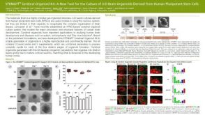 科学海报STEMdiff™ Cerebral Organoid Kit: A New Tool for the Culture of 3D Brain Organoids Derived from hPSCs


 沪公网安备31010102008431号