
 EasySep™小鼠TIL(CD45)正选试剂盒


技术资料
-

-
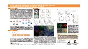 科学海报Efficient Generation of Lung Progenitor Cells From Human Pluripotent Stem Cells
Conference:
ISSCR 2021
-
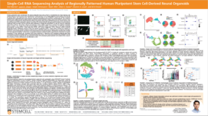 科学海报Single-Cell RNA Sequencing Analysis of Regionally Patterned Human Pluripotent Stem Cell-Derived Neural Organoids
Conference:
ISSCR 2021
-
-
-


 沪公网安备31010102008431号