Human (h) pluripotent stem cells (PSC) such as embryonic stem cells (ESC) can be directed into cardiomyocytes (CMs),representing a potential unlimited cell source for disease modeling,cardiotoxicity screening and myocardial repair. Although the electrophysiology of single hESC-CMs is now better defined,their multi-cellular arrhythmogenicity has not been thoroughly assessed due to the lack of a suitable experimental platform. Indeed,the generation of ventricular (V) fibrillation requires single-cell triggers as well as sustained multi-cellular reentrant events. Although native VCMs are aligned in a highly organized fashion such that electrical conduction is anisotropic for coordinated contractions,hESC-derived CM (hESC-CM) clusters are heterogenous and randomly organized,and therefore not representative of native conditions. Here,we reported that engineered alignment of hESC-VCMs on biomimetic grooves uniquely led to physiologically relevant responses. Aligned but not isotropic control preparations showed distinct longitudinal (L) and transverse (T) conduction velocities (CV),resembling the native human V anisotropic ratio (AR=LCV/TCV=1.8-2.0). Importantly,the total incidence of spontaneous and inducible arrhythmias significantly reduced from 57% in controls to 17-23% of aligned preparations,thereby providing a physiological baseline for assessing arrhythmogenicity. As such,promotion of pro-arrhythmic effect (e.g.,spatial dispersion by ?? adrenergic stimulation) could be better predicted. Mechanistically,such anisotropy-induced electrical stability was not due to maturation of the cellular properties of hESC-VCMs but their physical arrangement. In conclusion,not only do functional anisotropic hESC-VCMs engineered by multi-scale topography represent a more accurate model for efficacious drug discovery and development as well as arrhythmogenicity screening (of pharmacological and genetic factors),but our approach may also lead to future transplantable prototypes with improved efficacy and safety against arrhythmias. ?? 2013.
View Publication

 EasySep™小鼠TIL(CD45)正选试剂盒


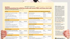 挂图Reporting Practices for Publishing Results with hPSCs Learn how to plan and conduct your human pluripotent stem cell (hPSC)-based research following the ISSCR’s Standards for Human Stem Cell Use in Research
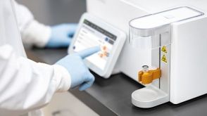 实验方案Optimizing Delivery Efficiency with Fluorescent Dextran Using the CellPore™ Transfection System


 沪公网安备31010102008431号