
 EasySep™小鼠TIL(CD45)正选试剂盒


技术资料
-
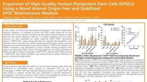 科学海报Expansion of High-Quality Human Pluripotent Stem Cells (hPSCs) Using a Novel Animal Origin-Free and Stabilized hPSC Maintenance Medium
Conference:
The New York Stem Cell Foundation Conference 2020
-

-

-

-
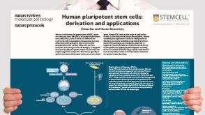 挂图Derivation and Applications of Human Pluripotent Stem Cells Overview of the derivation of human embryonic stem cells (hESCs) and induced pluripotent stem cells (iPSCs)
 -
 1:02:03
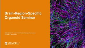
线上讲座Building Brain Organoids and AssemBloids™ to Study Human Development and Disease发布日期: 10/30/2020
1:02:03
线上讲座Building Brain Organoids and AssemBloids™ to Study Human Development and Disease发布日期: 10/30/2020 -


 沪公网安备31010102008431号