DiMascio L et al. (MAR 2007)
The Journal of Immunology 178 6 3511--3520
Identification of Adiponectin as a Novel Hemopoietic Stem Cell Growth Factor
The hemopoietic microenvironment consists of a diverse repertoire of cells capable of providing signals that influence hemopoietic stem cell function. Although the role of osteoblasts and vascular endothelial cells has recently been characterized,the function of the most abundant cell type in the bone marrow,the adipocyte,is less defined. Given the emergence of a growing number of adipokines,it is possible that these factors may also play a role in regulating hematopoiesis. Here,we investigated the role of adiponectin,a secreted molecule derived from adipocytes,in hemopoietic stem cell (HSC) function. We show that adiponectin is expressed by components of the HSC niche and its receptors AdipoR1 and AdipoR2 are expressed by HSCs. At a functional level,adiponectin influences HSCs by increasing their proliferation,while retaining the cells in a functionally immature state as determined by in vitro and in vivo assays. We also demonstrate that adiponectin signaling is required for optimal HSC proliferation both in vitro and in long term hemopoietic reconstitution in vivo. Finally we show that adiponectin stimulation activates p38 MAPK,and that inhibition of this pathway abrogates adiponectin's proliferative effect on HSCs. These studies collectively identify adiponectin as a novel regulator of HSC function and suggest that it acts through a p38 dependent pathway.
View Publication
产品号#:
03434
03444
72632
72634
产品名:
MethoCult™ GF M3434
MethoCult™ GF M3434
SB202190
SB202190
Brandl M et al. (AUG 1999)
Experimental hematology 27 8 1264--70
Bispecific antibody fragments with CD20 X CD28 specificity allow effective autologous and allogeneic T-cell activation against malignant cells in peripheral blood and bone marrow cultures from patients with B-cell lineage leukemia and lymphoma.
Bispecific antibodies directed against tumor-associated target antigens and to surface receptors mediating T-cell activation,such as the TCR/CD3 complex and the costimulatory receptor CD28,are capable of mediating T-cell activation resulting in tumor cell killing. In this study,we used the B-cell-associated antigens CD19 and CD20 as target structures on human leukemic cells. We found that a combination of bispecific antibody fragments (bsFab2) with target x CD3 and target x CD28 specificity induces vigorous autologous T-cell activation and killing of malignant cells in peripheral blood and bone marrow cultures from patients with chronic lymphocytic leukemia and follicular lymphoma. The bsFab2 targeting CD20 were considerably more effective than those binding to CD19. The colony-forming capacity of treated bone marrow was impaired due to large amounts of tumor necrosis factor alpha produced during bsFab2-induced T-cell activation. Neutralizing tumor necrosis factor alpha antibodies were found to reverse this negative effect without affecting T-cell activation and tumor cell killing. CD20 x CD28 bsFab2,when used alone rather than in combination,markedly improved the recognition of leukemic cells by allogeneic T cells. Therefore,these reagents may be capable of enhancing the immunogenicity of leukemic cells in general and,in particular,of increasing the antileukemic activity of allogeneic donor buffy coat cells in relapsed bone marrow transplanted patients.
View Publication
Sjogren A-KM et al. (MAY 2007)
The Journal of clinical investigation 117 5 1294--304
GGTase-I deficiency reduces tumor formation and improves survival in mice with K-RAS-induced lung cancer.
Protein geranylgeranyltransferase type I (GGTase-I) is responsible for the posttranslational lipidation of CAAX proteins such as RHOA,RAC1,and cell division cycle 42 (CDC42). Inhibition of GGTase-I has been suggested as a strategy to treat cancer and a host of other diseases. Although several GGTase-I inhibitors (GGTIs) have been synthesized,they have very different properties,and the effects of GGTIs and GGTase-I deficiency are unclear. One concern is that inhibiting GGTase-I might lead to severe toxicity. In this study,we determined the effects of GGTase-I deficiency on cell viability and K-RAS-induced cancer development in mice. Inactivating the gene for the critical beta subunit of GGTase-I eliminated GGTase-I activity,disrupted the actin cytoskeleton,reduced cell migration,and blocked the proliferation of fibroblasts expressing oncogenic K-RAS. Moreover,the absence of GGTase-I activity reduced lung tumor formation,eliminated myeloproliferative phenotypes,and increased survival of mice in which expression of oncogenic K-RAS was switched on in lung cells and myeloid cells. Interestingly,several cell types remained viable in the absence of GGTase-I,and myelopoiesis appeared to function normally. These findings suggest that inhibiting GGTase-I may be a useful strategy to treat K-RAS-induced malignancies.
View Publication
产品号#:
03234
产品名:
MethoCult™ M3234
Heuser M et al. (SEP 2007)
Blood 110 5 1639--47
MN1 overexpression induces acute myeloid leukemia in mice and predicts ATRA resistance in patients with AML.
Overexpression of wild-type MN1 is a negative prognostic factor in patients with acute myeloid leukemia (AML) with normal cytogenetics. We evaluated whether MN1 plays a functional role in leukemogenesis. We demonstrate using retroviral gene transfer and bone marrow (BM) transplantation that MN1 overexpression rapidly induces lethal AML in mice. Insertional mutagenesis and chromosomal instability were ruled out as secondary aberrations. MN1 increased resistance to all-trans retinoic acid (ATRA)-induced cell-cycle arrest and differentiation by more than 3000-fold in vitro. The differentiation block could be released by fusion of a transcriptional activator (VP16) to MN1 without affecting the ability to immortalize BM cells,suggesting that MN1 blocks differentiation by transcriptional repression. We then evaluated whether MN1 expression levels in patients with AML (excluding M3-AML) correlated with resistance to ATRA treatment in elderly patients uniformly treated within treatment protocol AMLHD98-B. Strikingly,patients with low MN1 expression who received ATRA had a significantly prolonged event-free (P = .008) and overall (P = .04) survival compared with patients with either low MN1 expression and no ATRA,or high MN1 expression with or without ATRA. MN1 is a unique oncogene in hematopoiesis that both promotes proliferation/self-renewal and blocks differentiation,and may become useful as a predictive marker in AML treatment.
View Publication
产品号#:
03234
产品名:
MethoCult™ M3234
Han X-D et al. (MAY 2007)
Proceedings of the National Academy of Sciences of the United States of America 104 21 9007--11
Fetal gene therapy of alpha-thalassemia in a mouse model.
Fetuses with homozygous alpha-thalassemia usually die at the third trimester of pregnancy or soon after birth. Hence,the disease could potentially be a target for fetal gene therapy. We have previously established a mouse model of alpha-thalassemia. These mice mimic the human alpha-thalassemic conditions and can be used as preclinical models for fetal gene therapy. We tested a lentiviral vector containing the HS 2,3,and 4 of the beta-LCR,a central polypurine tract element,and the beta-globin gene promoter directing either the EGFP or the human alpha-globin gene. We showed that the GFP expression was erythroid-specific and detected in BFU-E colonies and the erythroid progenies of CFU-GEMM. For in utero gene delivery,we did yolk sac vessel injection at midgestation of mouse embryos. The recipient mice were analyzed after birth for human alpha-globin gene expression. In the newborn,human alpha-globin gene expression was detected in the liver,spleen,and peripheral blood. The human alpha-globin gene expression was at the peak at 3-4 months,when it reached 20% in some recipients. However,the expression declined at 7 months. Colony-forming assays in these mice showed low abundance of the transduced human alpha-globin gene in their BFU-E and CFU-GEMM and the lack of its transcript. Thus,lentiviral vectors can be an effective vehicle for delivering the human alpha-globin gene into erythroid cells in utero,but,in the mouse model,delivery at late midgestation could not transduce hematopoietic stem cells adequately to sustain gene expression.
View Publication
产品号#:
03434
03444
产品名:
MethoCult™ GF M3434
MethoCult™ GF M3434
Dedhia PH et al. (AUG 2010)
Blood 116 8 1321--8
Differential ability of Tribbles family members to promote degradation of C/EBPalpha and induce acute myelogenous leukemia.
Trib1,Trib2,and Trib3 are mammalian homologs of Tribbles,an evolutionarily conserved Drosophila protein family that mediates protein degradation. Tribbles proteins function as adapters to recruit E3 ubiquitin ligases and enhance ubiquitylation of the target protein to promote its degradation. Increased Trib1 and Trib2 mRNA expression occurs in human myeloid leukemia and induces acute myeloid leukemia in mice,whereas Trib3 has not been associated with leukemia. Given the high degree of structural conservation among Tribbles family members,we directly compared the 3 mammalian Tribbles in hematopoietic cells by reconstituting mice with hematopoietic stem cells retrovirally expressing these proteins. All mice receiving Trib1 or Trib2 transduced hematopoietic stem cells developed acute myeloid leukemia,whereas Trib3 mice did not. Our previous data indicated that Trib2-mediated degradation of the transcription factor,CCAAT/enhancer-binding protein-alpha (C/EBPalpha),is important for leukemogenesis. Similar to Trib2,Trib1 induced C/EBPalpha degradation and inhibited its function. In contrast,Trib3 failed to inactivate or promote efficient degradation of C/EBPalpha. These data reveal that the 3 Tribbles homologs differ in their ability to promote degradation of C/EBPalpha,which account for their differential ability to induce leukemia.
View Publication
产品号#:
03231
产品名:
MethoCult™ M3231
Vanneaux V et al. (JAN 2010)
Cell transplantation 19 9 1143--55
In vitro and in vivo analysis of endothelial progenitor cells from cryopreserved umbilical cord blood: are we ready for clinical application?
Umbilical cord blood (CB) represents a main source of circulating endothelial progenitor cells (cEPCs). In view of their clinical use,in either the autologous or allogeneic setting,cEPCs should likely be expanded from CB kept frozen in CB banks. In this study,we compared the expansion,functional features,senescence pattern over culture,and in vivo angiogenic potential of cEPCs isolated from fresh or cryopreserved CB (cryoCB). cEPCs could be isolated in only 59% of cryoCB compared to 94% for fresh CB,while CB units were matched in terms of initial volume,nucleated and CD34(+) cell number. Moreover,the number of endothelial colony-forming cells was significantly decreased when using cryoCB. Once cEPCs culture was established,the proliferation,migration,tube formation,and acetylated-LDL uptake potentials were similar in both groups. In addition,cEPCs derived from cryoCB displayed the same senescence status and telomeres length as that of cEPCs derived from fresh CB. Karyotypic aberrations were found in cells obtained from both fresh and cryoCB. In vivo,in a hind limb ischemia murine model,cEPCs from fresh and cryoCB were equally efficient to induce neovascularization. Thus,cEPCs isolated from cryoCB exhibited similar properties to those of fresh CB in vitro and in vivo. However,the low frequency of cEPCs colony formation after cryopreservation shed light on the need for specific freezing conditions adapted to cEPCs in view of their future clinical use.
View Publication
产品号#:
15026
15066
产品名:
RosetteSep™人造血祖细胞富集抗体混合物
RosetteSep™人造血祖细胞富集抗体混合物
Dí et al. (DEC 2007)
Cardiovascular research 76 3 517--27
Plasticity of CD133+ cells: role in pulmonary vascular remodeling.
OBJECTIVE: Studies in pulmonary arteries (PA) of patients with chronic obstructive pulmonary disease (COPD) suggest that bone marrow-derived endothelial progenitor cells (CD133(+)) may infiltrate the intima and differentiate into smooth muscle cells (SMC). This study aimed to evaluate the plasticity of CD133(+) cells to differentiate into SMC and endothelial cells (EC) in both cell culture and human isolated PA. METHODS: Plasticity of granulocyte-colony stimulator factor (G-CSF)-mobilized peripheral blood CD133(+) cells was assessed in co-cultures with primary lines of human PA endothelial cells (PAEC) or SMC (PASMC) and in isolated human PA. We also evaluated if the phenotype of differentiated progenitor cells was acquired by fusion or differentiation. RESULTS: The in vitro studies demonstrated CD133(+) cells may acquire the morphology and phenotype of the cells they were co-cultured with. CD133(+) cells co-incubated with human isolated PA were able to migrate into the intima and differentiate into SMC. Progenitor cell differentiation was produced without fusion with mature cells. CONCLUSIONS: We provide evidence of plasticity of CD133(+) cells to differentiate into both endothelial cells and SMC,reinforcing the idea of their potential role in the remodeling process of PA in COPD. This process was conducted by transdifferentiation and not by cell fusion.
View Publication
产品号#:
产品名:
Qiu C et al. (FEB 2008)
Blood 111 4 2400--8
Globin switches in yolk sac-like primitive and fetal-like definitive red blood cells produced from human embryonic stem cells.
We have previously shown that coculture of human embryonic stem cells (hESCs) for 14 days with immortalized fetal hepatocytes yields CD34(+) cells that can be expanded in serum-free liquid culture into large numbers of megaloblastic nucleated erythroblasts resembling yolk sac-derived cells. We show here that these primitive erythroblasts undergo a switch in hemoglobin (Hb) composition during late terminal erythroid maturation with the basophilic erythroblasts expressing predominantly Hb Gower I (zeta(2)epsilon(2)) and the orthochromatic erythroblasts hemoglobin Gower II (alpha(2)epsilon(2)). This suggests that the switch from Hb Gower I to Hb Gower II,the first hemoglobin switch in humans is a maturation switch not a lineage switch. We also show that extending the coculture of the hESCs with immortalized fetal hepatocytes to 35 days yields CD34(+) cells that differentiate into more developmentally mature,fetal liver-like erythroblasts,that are smaller,express mostly fetal hemoglobin,and can enucleate. We conclude that hESC-derived erythropoiesis closely mimics early human development because the first 2 human hemoglobin switches are recapitulated,and because yolk sac-like and fetal liver-like cells are sequentially produced. Development of a method that yields erythroid cells with an adult phenotype remains necessary,because the most mature cells that can be produced with current systems express less than 2% adult beta-globin mRNA.
View Publication
产品号#:
09600
09650
18056
18056RF
产品名:
StemSpan™ SFEM
StemSpan™ SFEM
Li H et al. (AUG 2010)
Blood 116 7 1060--9
Repression of Id2 expression by Gfi-1 is required for B-cell and myeloid development.
The development of mature blood cells from hematopoietic stem cells requires coordinated activities of transcriptional networks. Transcriptional repressor growth factor independence 1 (Gfi-1) is required for the development of B cells,T cells,neutrophils,and for the maintenance of hematopoietic stem cell function. However,the mechanisms by which Gfi-1 regulates hematopoiesis and how Gfi-1 integrates into transcriptional networks remain unclear. Here,we provide evidence that Id2 is a transcriptional target of Gfi-1,and repression of Id2 by Gfi-1 is required for B-cell and myeloid development. Gfi-1 binds to 3 conserved regions in the Id2 promoter and represses Id2 promoter activity in transient reporter assays. Increased Id2 expression was observed in multipotent progenitors,myeloid progenitors,T-cell progenitors,and B-cell progenitors in Gfi-1(-/-) mice. Knockdown of Id2 expression or heterozygosity at the Id2 locus partially rescues the B-cell and myeloid development but not the T-cell development in Gfi-1(-/-) mice. These studies demonstrate a role of Id2 in mediating Gfi-1 functions in B-cell and myeloid development and provide a direct link between Gfi-1 and the B-cell transcriptional network by its ability to repress Id2 expression.
View Publication
产品号#:
03234
产品名:
MethoCult™ M3234
Ghiaur G et al. (APR 2008)
Blood 111 7 3313--21
Rac1 is essential for intraembryonic hematopoiesis and for the initial seeding of fetal liver with definitive hematopoietic progenitor cells.
Definitive hematopoietic stem and progenitor cells (HSCs/Ps) originating from the yolk sac and/or para-aorta-splanchno-pleura/aorta-gonad-mesonephros are hypothesized to colonize the fetal liver,but mechanisms involved are poorly defined. The Rac subfamily of Rho GTPases has been shown to play essential roles in HSC/P localization to the bone marrow following transplantation. Here,we study the role of Rac1 in HSC/P migration during ontogeny and seeding of fetal liver. Using a triple-transgenic approach,we have deleted Rac1 in HSCs/Ps during very early embryonic development. Without Rac1,there was a decrease in circulating HSCs/Ps in the blood of embryonic day (E) 10.5 embryos,while yolk sac definitive hematopoiesis was quantitatively normal. Intraembryonic hematopoiesis was significantly impaired in Rac1-deficient embryos,culminating with absence of intra-aortic clusters and fetal liver hematopoiesis. At E10.5,Rac1-deficient HSCs/Ps displayed decreased transwell migration and impaired inter-action with the microenvironment in migration-dependent assays. These data suggest that Rac1 plays an important role in HSC/P migration during embryonic development and is essential for the emergence of intraembryonic hematopoiesis.
View Publication

 EasySep™小鼠TIL(CD45)正选试剂盒


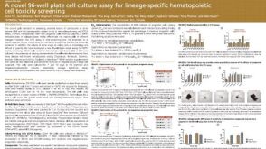 科学海报A Novel 96-well Plate Cell Culture Assay for Lineage-Specific Hematopoietic Cell Toxicity Screening
 沪公网安备31010102008431号