Moreau-Gaudry F et al. (NOV 2001)
Blood 98 9 2664--72
High-level erythroid-specific gene expression in primary human and murine hematopoietic cells with self-inactivating lentiviral vectors.
Use of oncoretroviral vectors in gene therapy for hemoglobinopathies has been impeded by low titer vectors,genetic instability,and poor expression. Fifteen self- inactivating (SIN) lentiviral vectors using 4 erythroid promoters in combination with 4 erythroid enhancers with or without the woodchuck hepatitis virus postregulatory element (WPRE) were generated using the enhanced green fluorescent protein as a reporter gene. Vectors with high erythroid-specific expression in cell lines were tested in primary human CD34(+) cells and in vivo in the murine bone marrow (BM) transplantation model. Vectors containing the ankyrin-1 promoter showed high-level expression and stable proviral transmission. Two vectors containing the ankyrin-1 promoter and 2 erythroid enhancers (HS-40 plus GATA-1 or HS-40 plus 5-aminolevulinate synthase intron 8 [I8] enhancers) and WPRE expressed at levels higher than the HS2/beta-promoter vector in bulk unilineage erythroid cultures and individual erythroid blast-forming units derived from human BM CD34(+) cells. Sca1(+)/lineage(-) Ly5.1 mouse hematopoietic cells,transduced with these 2 ankyrin-1 promoter vectors,were injected into lethally irradiated Ly5.2 recipients. Eleven weeks after transplantation,high-level expression was seen from both vectors in blood (63%-89% of red blood cells) and erythroid cells in BM (70%-86% engraftment),compared with negligible expression in myeloid and lymphoid lineages in blood,BM,spleen,and thymus (0%-4%). The I8/HS-40-containing vector encoding a hybrid human beta/gamma-globin gene led to 43% to 113% human gamma-globin expression/copy of the mouse alpha-globin gene. Thus,modular use of erythroid-specific enhancers/promoters and WPRE in SIN-lentiviral vectors led to identification of high-titer,stably transmitted vectors with high-level erythroid-specific expression for gene therapy of red cell diseases.
View Publication
产品号#:
产品名:
Baba Y et al. (AUG 2006)
Journal of immunology (Baltimore,Md. : 1950) 177 4 2294--303
Constitutively active beta-catenin promotes expansion of multipotent hematopoietic progenitors in culture.
This study was designed to investigate one component of the Wnt/beta-catenin signaling pathway that has been implicated in stem cell self-renewal. Retroviral-mediated introduction of stable beta-catenin to primitive murine bone marrow cells allowed the expansion of multipotential c-Kit(low)Sca-1(low/-)CD19(-) CD11b/Mac-1(-)Flk-2(-)CD43(+)AA4.1(+)NK1.1(-)CD3(-)CD11c(-)Gr-1(-)CD45R/B220(+) cells in the presence of stromal cells and cytokines. They generated myeloid,T,and B lineage lymphoid cells in culture,but had no T lymphopoietic potential when transplanted. Stem cell factor and IL-6 were found to be minimal requirements for long-term,stromal-free propagation,and a beta-catenin-transduced cell line was maintained for 5 mo with these defined conditions. Although multipotential and responsive to many normal stimuli in culture,it was unable to engraft several types of irradiated recipients. These findings support previous studies that have implicated the canonical Wnt pathway signaling in regulation of multipotent progenitors. In addition,we demonstrate how it may be experimentally manipulated to generate valuable cell lines.
View Publication
产品号#:
03434
03444
产品名:
MethoCult™ GF M3434
MethoCult™ GF M3434
Karp JE et al. (MAY 2009)
Blood 113 20 4841--52
Active oral regimen for elderly adults with newly diagnosed acute myelogenous leukemia: a preclinical and phase 1 trial of the farnesyltransferase inhibitor tipifarnib (R115777, Zarnestra) combined with etoposide.
The farnesyltransferase inhibitor tipifarnib exhibits modest activity against acute myelogenous leukemia. To build on these results,we examined the effect of combining tipifarnib with other agents. Tipifarnib inhibited signaling downstream of the farnesylated small G protein Rheb and synergistically enhanced etoposide-induced antiproliferative effects in lymphohematopoietic cell lines and acute myelogenous leukemia isolates. We subsequently conducted a phase 1 trial of tipifarnib plus etoposide in adults over 70 years of age who were not candidates for conventional therapy. A total of 84 patients (median age,77 years) received 224 cycles of oral tipifarnib (300-600 mg twice daily for 14 or 21 days) plus oral etoposide (100-200 mg daily on days 1-3 and 8-10). Dose-limiting toxicities occurred with 21-day tipifarnib. Complete remissions were achieved in 16 of 54 (30%) receiving 14-day tipifarnib versus 5 of 30 (17%) receiving 21-day tipifarnib. Complete remissions occurred in 50% of two 14-day tipifarnib cohorts: 3A (tipifarnib 600,etoposide 100) and 8A (tipifarnib 400,etoposide 200). In vivo,tipifarnib plus etoposide decreased ribosomal S6 protein phosphorylation and increased histone H2AX phosphorylation and apoptosis. Tipifarnib plus etoposide is a promising orally bioavailable regimen that warrants further evaluation in elderly adults who are not candidates for conventional induction chemotherapy. These clinical studies are registered at www.clinicaltrials.gov as NCT00112853.
View Publication
产品号#:
04434
04444
产品名:
MethoCult™ H4434 Classic
MethoCult™ H4434 Classic
Zhao Z et al. (JUL 2010)
Genes & development 24 13 1389--402
p53 loss promotes acute myeloid leukemia by enabling aberrant self-renewal.
The p53 tumor suppressor limits proliferation in response to cellular stress through several mechanisms. Here,we test whether the recently described ability of p53 to limit stem cell self-renewal suppresses tumorigenesis in acute myeloid leukemia (AML),an aggressive cancer in which p53 mutations are associated with drug resistance and adverse outcome. Our approach combined mosaic mouse models,Cre-lox technology,and in vivo RNAi to disable p53 and simultaneously activate endogenous Kras(G12D)-a common AML lesion that promotes proliferation but not self-renewal. We show that p53 inactivation strongly cooperates with oncogenic Kras(G12D) to induce aggressive AML,while both lesions on their own induce T-cell malignancies with long latency. This synergy is based on a pivotal role of p53 in limiting aberrant self-renewal of myeloid progenitor cells,such that loss of p53 counters the deleterious effects of oncogenic Kras on these cells and enables them to self-renew indefinitely. Consequently,myeloid progenitor cells expressing oncogenic Kras and lacking p53 become leukemia-initiating cells,resembling cancer stem cells capable of maintaining AML in vivo. Our results establish an efficient new strategy for interrogating oncogene cooperation,and provide strong evidence that the ability of p53 to limit aberrant self-renewal contributes to its tumor suppressor activity.
View Publication
产品号#:
03534
09600
09650
产品名:
MethoCult™ GF M3534
StemSpan™ SFEM
StemSpan™ SFEM
Ragu C et al. (NOV 2010)
Blood 116 22 4464--73
The transcription factor Srf regulates hematopoietic stem cell adhesion.
Adhesion properties of hematopoietic stem cells (HSCs) in the bone marrow (BM) niches control their migration and affect their cell-cycle dynamics. The serum response factor (Srf) regulates growth factor-inducible genes and genes controlling cytoskeleton structures involved in cell spreading,adhesion,and migration. We identified a role for Srf in HSC adhesion and steady-state hematopoiesis. Conditional deletion of Srf in BM cells resulted in a 3-fold expansion of the long- and short-term HSCs and multipotent progenitors (MPPs),which occurs without long-term modification of cell-cycle dynamics. Early differentiation steps to myeloid and lymphoid lineages were normal,but Srf loss results in alterations in mature-cell production and severe thrombocytopenia. Srf-null BM cells also displayed compromised engraftment properties in transplantation assays. Gene expression analysis identified Srf target genes expressed in HSCs,including a network of genes associated with cell migration and adhesion. Srf-null stem cells and MPPs displayed impair expression of the integrin network and decreased adherence in vitro. In addition,Srf-null mice showed increase numbers of circulating stem and progenitor cells,which likely reflect their reduced retention in the BM. Altogether,our results demonstrate that Srf is an essential regulator of stem cells and MPP adhesion,and suggest that Srf acts mainly through cell-matrix interactions and integrin signaling.
View Publication
产品号#:
03434
03444
09600
09650
产品名:
MethoCult™ GF M3434
MethoCult™ GF M3434
StemSpan™ SFEM
StemSpan™ SFEM
Mahdipour E et al. (JAN 2011)
Blood 117 3 815--26
Hoxa3 promotes the differentiation of hematopoietic progenitor cells into proangiogenic Gr-1+CD11b+ myeloid cells.
Injury induces the recruitment of bone marrow-derived cells (BMDCs) that contribute to the repair and regeneration process. The behavior of BMDCs in injured tissue has a profound effect on repair,but the regulation of BMDC behavior is poorly understood. Aberrant recruitment/retention of these cells in wounds of diabetic patients and animal models is associated with chronic inflammation and impaired healing. BMD Gr-1(+)CD11b(+) cells function as immune suppressor cells and contribute significantly to tumor-induced neovascularization. Here we report that Gr-1(+)CD11b(+) cells also contribute to injury-induced neovascularization,but show altered recruitment/retention kinetics in the diabetic environment. Moreover,diabetic-derived Gr-1(+)CD11b(+) cells fail to stimulate neovascularization in vivo and have aberrant proliferative,chemotaxis,adhesion,and differentiation potential. Previously we demonstrated that gene transfer of HOXA3 to wounds of diabetic mice is taken up by and expressed by recruited BMDCs. This is associated with a suppressed inflammatory response,enhanced neovascularization,and accelerated wound healing. Here we show that sustained expression of Hoxa3 in diabetic-derived BMD Gr-1(+)CD11b(+) cells reverses their diabetic phenotype. These findings demonstrate that manipulation of adult stem/progenitor cells ex vivo could be used as a potential therapy in patients with impaired wound healing.
View Publication

 EasySep™小鼠TIL(CD45)正选试剂盒


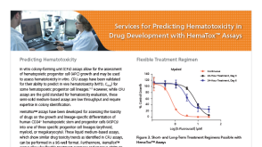 产品手册Services for Predicting Hematotoxicity in Drug Development with HemaTox™ Assays


 沪公网安备31010102008431号