
 EasySep™小鼠TIL(CD45)正选试剂盒


技术资料
-
产品号#:
19853
19853RF
17961
17961RF
产品名:
EasySep™小鼠CD8+ T细胞分选试剂盒
RoboSep™ 小鼠CD8+ T细胞分选试剂盒
EasySep™人Naïve Pan-T细胞分选试剂盒
RoboSep™ 人Naïve Pan-T细胞分选试剂盒
-
产品号#:
17877
17877RF
产品名:
EasySep™人CD138正选试剂盒 II
RoboSep™ 人CD138正选试剂盒 II
-
产品号#:
17952
17952RF
100-0696
产品名:
EasySep™人CD4+ T细胞分选试剂盒
RoboSep™ 人CD4+ T细胞分选试剂盒
EasySep™人CD4+ T细胞分离试剂盒
-
产品号#:
17853
17853RF
100-0699
产品名:
EasySep™人CD8正选试剂盒 II
RoboSep™ 人CD8正选试剂盒 II
EasySep™人CD8阳性选择试剂盒II
-
 1:07:14
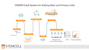
线上讲座Optimized Workflows for High-Efficiency Genome Editing in Stem and Primary Cell Types发布日期: 09/09/2019
1:07:14
线上讲座Optimized Workflows for High-Efficiency Genome Editing in Stem and Primary Cell Types发布日期: 09/09/2019 -
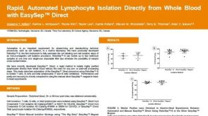 科学海报Rapid, Automated Lymphocyte Isolation Directly from Whole Blood with EasySep™ Direct
Conference:
EFI 2019
-
-
-
-

 沪公网安备31010102008431号