Krummen M et al. (JUL 2010)
Journal of leukocyte biology 88 1 189--99
Release of IL-12 by dendritic cells activated by TLR ligation is dependent on MyD88 signaling, whereas TRIF signaling is indispensable for TLR synergy.
Recently,it has been shown that certain combinations of TLR ligands act in synergy to induce the release of IL-12 by DCs. In this study,we sought to define the critical parameters underlying TLR synergy. Our data show that TLR ligands act synergistically if MyD88- and TRIF-dependent ligands are combined. TLR4 uses both of these adaptor molecules,thus activation via TLR4 proved to be a synergistic event on its own. TLR synergy did not affect all aspects of DC activation but enhanced primarily the release of certain cytokines,particularly IL-12,whereas the expression of costimulatory molecules remained unchanged. Consequently,synergistic activation of DC did not affect their ability to induce T cell proliferation but resulted in T(H)1-biased responses in vitro and in vivo. Furthermore,we examined the impact of TLR ligand combinations on primary DC in vitro but observed only modest effects with a combination of CpG + Poly (I:C). However,noticeable synergy in terms of IL-12 production by DCs was detectable in vivo after systemic administration of CpG + Poly (I:C). Finally,we show that synergy is partially dependent on IFNAR signaling but does not require the release of IFNs to the enviroment,suggesting an autocrine action of type I IFNs.
View Publication
产品号#:
18752
18752RF
21000
20119
20155
18758
18758RF
18768
18768RF
产品名:
RoboSep™- S
RoboSep™ 吸头组件抛光剂
RoboSep™分选管套装(9个塑料管)
Harry RA et al. (NOV 2010)
Annals of the rheumatic diseases 69 11 2042--50
Generation and characterisation of therapeutic tolerogenic dendritic cells for rheumatoid arthritis.
OBJECTIVES: Tolerogenic dendritic cells (tolDCs) constitute a promising experimental treatment for targeting autoreactive T cells in autoimmune diseases,including rheumatoid arthritis (RA). The authors' goal is to bring tolDC therapy for RA to the clinic. Here the authors address key translational issues related to the manufacturing of tolDCs from RA patients with current good manufacturing practice (cGMP)-compliant reagents,the stability of tolDCs,and the selection of suitable quality control markers. METHODS: Human monocyte-derived tolDCs were established from RA patients and healthy controls (HCs) using the immunosuppressive drugs dexamethasone and vitamin D₃,and the cGMP-grade immunomodulator,monophosphoryl lipid A,in the cGMP-compliant medium,CellGroDC. The functionality of tolDCs and tolDC-modulated autologous CD4 T cells was determined by flow cytometry,[³H]thymidine incorporation and ELISA. RESULTS: Clinical-grade tolDCs established from patients with RA exhibit a typical tolerogenic phenotype of reduced costimulatory molecules,low production of proinflammatory cytokines and impaired stimulation of autologous antigen-specific T cells,comparable to HC tolDCs. Toll-like receptor 2 (TLR-2) was highly expressed by tolDCs but not mature DCs. Furthermore,tolDCs suppressed mature DC-induced T cell proliferation,interferon γ and interleukin 17 production,and rendered T cells hyporesponsive to further stimulation. Importantly,tolDCs were phenotypically stable in the absence of immunosuppressive drugs and were refractory to further challenge with proinflammatory mediators. CONCLUSIONS: tolDCs established from patients with RA are comparable to those derived from healthy donors. TLR-2 was identified as an ideal marker for quality control of tolDCs. Potently tolerogenic and highly stable,these tolDCs are a promising cellular therapeutic for tailored immunomodulation in the treatment of RA.
View Publication
产品号#:
15022
15062
产品名:
RosetteSep™人CD4+ T细胞富集抗体混合物
RosetteSep™人CD4+ T细胞富集抗体混合物
Imbert A-M et al. (OCT 2006)
Blood 108 8 2578--86
CD99 expressed on human mobilized peripheral blood CD34+ cells is involved in transendothelial migration.
Hematopoietic progenitor cell trafficking is an important phenomenon throughout life. It is thought to occur in sequential steps,similar to what has been described for mature leukocytes. Molecular actors have been identified for each step of leukocyte migration; recently,CD99 was shown to play a part during transendothelial migration. We explored the expression and role of CD99 on human hematopoietic progenitors. We demonstrate that (1) CD34+ cells express CD99,albeit with various intensities; (2) subsets of CD34+ cells with high or low levels of CD99 expression produce different numbers of erythroid,natural killer (NK),or dendritic cells in the in vitro differentiation assays; (3) the level of CD99 expression is related to the ability to differentiate toward B cells; (4) CD34+ cells that migrate through an endothelial monolayer in response to SDF-1alpha and SCF display the highest level of CD99 expression; (5) binding of a neutralizing antibody to CD99 partially inhibits transendothelial migration of CD34+ progenitors in an in vitro assay; and (6) binding of a neutralizing antibody to CD99 reduces homing of CD34+ progenitors xenotransplanted in NOD-SCID mice. We conclude that expression of CD99 on human CD34+ progenitors has functional significance and that CD99 may be involved in transendothelial migration of progenitors.
View Publication
产品号#:
01700
01705
04230
01702
产品名:
ALDEFLUOR™ 试剂盒
ALDEFLUOR™ DEAB试剂, 1.5 mM, 1 mL
MethoCult™ H4230
ALDEFLUOR™检测缓冲液
Marzaioli V et al. ( 2017)
Blood 130 15 1734--1745
NOX5 and p22phox are 2 novel regulators of human monocytic differentiation into dendritic cells.
Dendritic cells (DCs) are a heterogeneous population of professional antigen-presenting cells and are key cells of the immune system,acquiring different phenotypes in accordance with their localization during the immune response. A subset of inflammatory DCs is derived from circulating monocytes (Mo) and has a key role in inflammation and infection. The pathways controlling Mo-DC differentiation are not fully understood. Our objective was to investigate the possible role of nicotinamide adenine dinucleotide phosphate reduced form oxidases (NOXs) in Mo-DC differentiation. In this study,we revealed that Mo-DC differentiation was inhibited by NOX inhibitors and reactive oxygen species scavengers. We show that the Mo-DC differentiation was dependent on p22phox,and not on gp91phox/NOX2,as shown by the reduced Mo-DC differentiation observed in chronic granulomatous disease patients lacking p22phox. Moreover,we revealed that NOX5 expression was strongly increased during Mo-DC differentiation,but not during Mo-macrophage differentiation. NOX5 was expressed in circulating myeloid DC,and at a lower level in plasmacytoid DC. Interestingly,NOX5 was localized at the outer membrane of the mitochondria and interacted with p22phox in Mo-DC. Selective inhibitors and small interfering RNAs for NOX5 indicated that NOX5 controlled Mo-DC differentiation by regulating the JAK/STAT/MAPK and NFκB pathways. These data demonstrate that the NOX5-p22phox complex drives Mo-DC differentiation,and thus could be critical for immunity and inflammation.
View Publication
产品号#:
19061
19061RF
19062
19062RF
19359
19359RF
100-0697
产品名:
EasySep™人髓样DC富集试剂盒
RoboSep™ 人髓样DC富集试剂盒
EasySep™人浆细胞样DC富集试剂盒
RoboSep™ 人浆细胞样DC富集试剂盒含滤芯吸头
EasySep™人单核细胞分选试剂盒
RoboSep™ 人单核细胞分选试剂盒
EasySep™人单核细胞分选试剂盒
Xu MM et al. (AUG 2017)
Immunity 47 2 363--373.e5
Dendritic Cells but Not Macrophages Sense Tumor Mitochondrial DNA for Cross-priming through Signal Regulatory Protein α Signaling.
Inhibition of cytosolic DNA sensing represents a strategy that tumor cells use for immune evasion,but the underlying mechanisms are unclear. Here we have shown that CD47-signal regulatory protein α (SIRPα) axis dictates the fate of ingested DNA in DCs for immune evasion. Although macrophages were more potent in uptaking tumor DNA,increase of DNA sensing by blocking the interaction of SIRPα with CD47 preferentially occurred in dendritic cells (DCs) but not in macrophages. Mechanistically,CD47 blockade enabled the activation of NADPH oxidase NOX2 in DCs,which in turn inhibited phagosomal acidification and reduced the degradation of tumor mitochondrial DNA (mtDNA) in DCs. mtDNA was recognized by cyclic-GMP-AMP synthase (cGAS) in the DC cytosol,contributing to type I interferon (IFN) production and antitumor adaptive immunity. Thus,our findings have demonstrated how tumor cells inhibit innate sensing in DCs and suggested that the CD47-SIRPα axis is critical for DC-driven antitumor immunity.
View Publication
产品号#:
18780
18780RF
18781
18781RF
19853
19853RF
70025
70025.1
70025.2
70025.3
产品名:
EasySep™小鼠CD11c正选试剂盒II
RoboSep™ 小鼠CD11c正选试剂盒II
EasySep™小鼠CD11c正选试剂盒II及脾脏解离液
RoboSep™ 小鼠CD11c正选试剂盒II及脾脏解离液
EasySep™小鼠CD8+ T细胞分选试剂盒
RoboSep™ 小鼠CD8+ T细胞分选试剂盒
冻存的人外周血单个核细胞
冻存的人外周血单个核细胞
冻存的人外周血单个核细胞
冻存的人外周血单个核细胞
Schulz O et al. (DEC 2009)
The Journal of experimental medicine 206 13 3101--14
Intestinal CD103+, but not CX3CR1+, antigen sampling cells migrate in lymph and serve classical dendritic cell functions.
Chemokine receptor CX3CR1(+) dendritic cells (DCs) have been suggested to sample intestinal antigens by extending transepithelial dendrites into the gut lumen. Other studies identified CD103(+) DCs in the mucosa,which,through their ability to synthesize retinoic acid (RA),appear to be capable of generating typical signatures of intestinal adaptive immune responses. We report that CD103 and CX3CR1 phenotypically and functionally characterize distinct subsets of lamina propria cells. In contrast to CD103(+) DC,CX3CR1(+) cells represent a nonmigratory gut-resident population with slow turnover rates and poor responses to FLT-3L and granulocyte/macrophage colony-stimulating factor. Direct visualization of cells in lymph vessels and flow cytometry of mouse intestinal lymph revealed that CD103(+) DCs,but not CX3CR1-expressing cells,migrate into the gut draining mesenteric lymph nodes (LNs) under steady-state and inflammatory conditions. Moreover,CX3CR1(+) cells displayed poor T cell stimulatory capacity in vitro and in vivo after direct injection of cells into intestinal lymphatics and appeared to be less efficient at generating RA compared with CD103(+) DC. These findings indicate that selectively CD103(+) DCs serve classical DC functions and initiate adaptive immune responses in local LNs,whereas CX3CR1(+) populations might modulate immune responses directly in the mucosa and serve as first line barrier against invading enteropathogens.
View Publication
产品号#:
01700
01705
01701
01702
产品名:
ALDEFLUOR™ 试剂盒
ALDEFLUOR™ DEAB试剂, 1.5 mM, 1 mL
ALDEFLUOR™检测缓冲液
Megjugorac NJ et al. (MAY 2010)
Blood 115 21 4185--90
IL-4 enhances IFN-lambda1 (IL-29) production by plasmacytoid DCs via monocyte secretion of IL-1Ra.
The type-III interferon (IFN) family is composed of 3 molecules in humans: IFN-lambda1 (interleukin-29 [IL-29]),IFN-lambda2 (IL-28A),and IFN-lambda3 (IL-28B),each of which signals through the same receptor complex. Plasmacytoid dendritic cells (pDCs) are major IFN-lambda producers among peripheral lymphocytes. Recently,it has been shown that IFN-lambda1 exerts a powerful inhibitory effect over the T-helper 2 (Th2) response by antagonizing the effect of IL-4 on CD4(+) T cells and inhibiting the production of Th2-associated cytokines. Here,we asked whether Th2 cytokines exert reciprocal control over IFN-lambda production. IL-4 treatment during stimulation of human peripheral lymphocytes significantly elevated IFN-lambda1 transcription and secretion. However,pDCs were not directly responsive to IL-4. Using depletion and reconstitution experiments,we showed that IL-4-responsive monocytes are an intermediary cell,responding to IL-4 by elevating their secretion of IL-1 receptor antagonist (IL-Ra); this IL-1Ra acts on pDCs to elevate their IFN-lambda1 output. Thus,our experiments revealed a novel mechanism for regulation of both IFN-lambda1 production and pDC function,and suggests an expanded immunomodulatory role for Th2-associated cytokines.
View Publication
产品号#:
18051
18051RF
18054
18054RF
18055
18055RF
18058
18058RF
19062
19062RF
19058
19058RF
18551
18551RF
18561
100-1525
产品名:
EasySep™人浆细胞样DC富集试剂盒
RoboSep™ 人浆细胞样DC富集试剂盒含滤芯吸头
EasySep™人单核细胞富集试剂盒(不去除CD16)
RoboSep™ 人单核细胞富集试剂盒(不去除CD16)含滤芯吸头
EasySep™人单核细胞富集试剂盒(不去除CD16)
Onai N et al. (JAN 2006)
The Journal of experimental medicine 203 1 227--38
Activation of the Flt3 signal transduction cascade rescues and enhances type I interferon-producing and dendritic cell development.
Flt3 ligand (Flt3L) is a nonredundant cytokine in type I interferon-producing cell (IPC) and dendritic cell (DC) development,and IPC and DC differentiation potential is confined to Flt3+ hematopoietic progenitor cells. Here,we show that overexpression of human Flt3 in Flt3- (Flt3(-)Lin(-)IL-7Ralpha(-)Thy1.1(-)c-Kit+) and Flt3+ (Flt3(+)Lin(-)IL-7Ralpha(-)Thy1.1(-)c-Kit+) hematopoietic progenitors rescues and enhances their IPC and DC differentiation potential,respectively. In defined hematopoietic cell populations,such as Flt3- megakaryocyte/erythrocyte-restricted progenitors (MEPs),enforced Flt3 signaling induces transcription of IPC,DC,and granulocyte/macrophage (GM) development-affiliated genes,including STAT3,PU.1,and G-/M-/GM-CSFR,and activates differentiation capacities to these lineages. Moreover,ectopic expression of Flt3 downstream transcription factors STAT3 or PU.1 in Flt3- MEPs evokes Flt3 receptor expression and instructs differentiation into IPCs,DCs,and myelomonocytic cells,whereas GATA-1 expression and consecutive megakaryocyte/erythrocyte development is suppressed. Based on these data,we propose a demand-regulated,cytokine-driven DC and IPC regeneration model,in which high Flt3L levels initiate a self-sustaining,Flt3-STAT3- and Flt3-PU.1-mediated IPC and DC differentiation program in Flt3+ hematopoietic progenitor cells.
View Publication
产品号#:
04100
产品名:
MethoCult™ H4100
Pereira RC et al. ( 2016)
Frontiers in immunology 7 415
Human Articular Chondrocytes Regulate Immune Response by Affecting Directly T Cell Proliferation and Indirectly Inhibiting Monocyte Differentiation to Professional Antigen-Presenting Cells.
Autologous chondrocyte implantation is the current gold standard cell therapy for cartilage lesions. However,in some instances,the heavily compromised health of the patient can either impair or limit the recovery of the autologous chondrocytes and a satisfactory outcome of the implant. Allogeneic human articular chondrocytes (hAC) could be a good alternative,but the possible immunological incompatibility between recipient and hAC donor should be considered. Herein,we report that allogeneic hAC inhibited T lymphocyte response to antigen-dependent and -independent proliferative stimuli. This effect was maximal when T cells and hAC were in contact and it was not relieved by the addition of exogenous lymphocyte growth factor interleukin (IL)-2. More important,hAC impaired the differentiation of peripheral blood monocytes induced with granulocyte monocyte colony-stimulating factor and IL-4 (Mo) to professional antigen-presenting cells,such as dendritic cells (DC). Indeed,a marked inhibition of the onset of the CD1a expression and an ineffective downregulation of CD14 antigens was observed in Mo-hAC co-cultures. Furthermore,compared to immature or mature DC,Mo from Mo-hAC co-cultures did not trigger an efficacious allo-response. The prostaglandin (PG) E2 present in the Mo-hAC co-culture conditioned media is a putative candidate of the hAC-mediated inhibition of Mo maturation. Altogether,these findings indicate that allogeneic hAC inhibit,rather than trigger,immune response and strongly suggest that an efficient chondrocyte implantation could be possible also in an allogeneic setting.
View Publication
产品号#:
17951
17951RF
17952
17952RF
18099
18099RF
100-0695
100-0696
产品名:
EasySep™人T细胞分选试剂盒
RoboSep™ 人T细胞分选试剂盒
EasySep™人CD4+ T细胞分选试剂盒
RoboSep™ 人CD4+ T细胞分选试剂盒
EasySep™人T细胞分选试剂盒
EasySep™人CD4+ T细胞分离试剂盒
G. B. Cross et al. ( 2019)
PloS one 14 5 e0216616
Impact of selective immune-cell depletion on growth of Mycobacterium tuberculosis (Mtb) in a whole-blood bactericidal activity (WBA) assay.
We investigated the contribution of host immune cells to bacterial killing in a whole-blood bactericidal activity (WBA) assay,an ex vivo model used to test efficacy of drugs against mycobacterium tuberculosis (Mtb). We performed WBA assays with immuno-magnetic depletion of specific cell types,in the presence or absence of rifampicin. Innate immune cells decreased Mtb growth in absence of drug,but appeared to diminish the cidal activity of rifampicin,possibly attributable to intracellular bacterial sequestration. Adaptive immune cells had no effect with or without drug. The WBA assay may have potential for testing adjunctive host-directed therapies acting on phagocytic cells.
View Publication
产品号#:
18000
产品名:
EasySep™磁极
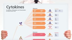
挂图
Human Immune Cytokines
Infographic of key cytokines for expansion, differentiation and characterization of major immune cell types

 EasySep™小鼠TIL(CD45)正选试剂盒


 挂图Human Immune Cytokines Infographic of key cytokines for expansion, differentiation and characterization of major immune cell types

 沪公网安备31010102008431号