Figueroa G et al. (OCT 2016)
Journal of visualized experiments : JoVE 116
Characterization of Human Monocyte-derived Dendritic Cells by Imaging Flow Cytometry: A Comparison between Two Monocyte Isolation Protocols.
Dendritic cells (DCs) are antigen presenting cells of the immune system that play a crucial role in lymphocyte responses,host defense mechanisms,and pathogenesis of inflammation. Isolation and study of DCs have been important in biological research because of their distinctive features. Although they are essential key mediators of the immune system,DCs are very rare in blood,accounting for approximately 0.1 - 1% of total blood mononuclear cells. Therefore,alternatives for isolation methods rely on the differentiation of DCs from monocytes isolated from peripheral blood mononuclear cells (PBMCs). The utilization of proper isolation techniques that combine simplicity,affordability,high purity,and high yield of cells is imperative to consider. In the current study,two distinct methods for the generation of DCs will be compared. Monocytes were selected by adherence or negatively enriched using magnetic separation procedure followed by differentiation into DCs with IL-4 and GM-CSF. Monocyte and MDDC viability,proliferation,and phenotype were assessed using viability dyes,MTT assay,and CD11c/ CD14 surface marker analysis by imaging flow cytometry. Although the magnetic separation method yielded a significant higher percentage of monocytes with higher proliferative capacity when compared to the adhesion method,the findings have demonstrated the ability of both techniques to simultaneously generate monocytes that are capable of proliferating and differentiating into viable CD11c+ MDDCs after seven days in culture. Both methods yielded textgreater 70% CD11c+ MDDCs. Therefore,our results provide insights that contribute to the development of reliable methods for isolation and characterization of human DCs.
View Publication
产品号#:
19059
19059RF
产品名:
EasySep™人单核细胞富集试剂盒
RoboSep™ 人单核细胞富集试剂盒含滤芯吸头
Tyagi RK et al. (FEB 2017)
Scientific reports 7 41083
Human IDO-competent, long-lived immunoregulatory dendritic cells induced by intracellular pathogen, and their fate in humanized mice.
Targeting of myeloid-dendritic cell receptor DC-SIGN by numerous chronic infectious agents,including Porphyromonas gingivalis,is shown to drive-differentiation of monocytes into dysfunctional mDCs. These mDCs exhibit alterations of their fine-tuned homeostatic function and contribute to dysregulated immune-responses. Here,we utilize P. gingivalis mutant strains to show that pathogen-differentiated mDCs from primary human-monocytes display anti-apoptotic profile,exhibited by elevated phosphorylated-Foxo1,phosphorylated-Akt1,and decreased Bim-expression. This results in an overall inhibition of DC-apoptosis. Direct stimulation of complex component CD40 on DCs leads to activation of Akt1,suggesting CD40 involvement in anti-apoptotic effects observed. Further,these DCs drove dampened CD8(+) T-cell and Th1/Th17 effector-responses while inducing CD25(+)Foxp3(+)CD127(-) Tregs. In vitro Treg induction was mediated by DC expression of indoleamine 2,3-dioxygenase,and was confirmed in IDO-KO mouse model. Pathogen-infected &CMFDA-labeled MoDCs long-lasting survival was confirmed in a huMoDC reconstituted humanized mice. In conclusion,our data implicate PDDCs as an important target for resolution of chronic infection.
View Publication
产品号#:
17858
17858RF
18058
18058RF
15028
15068
15628
15668
100-0694
产品名:
EasySep™人CD14正选试剂盒II
RoboSep™ 人CD14正选试剂盒II
RosetteSep™人单核细胞富集抗体混合物
RosetteSep™人单核细胞富集抗体混合物
RosetteSep™人单核细胞去除抗体混合物
RosetteSep™人单核细胞去除抗体混合物
EasySep™人CD14正选试剂盒II
O'Mahony L et al. (APR 2006)
American journal of physiology. Gastrointestinal and liver physiology 290 4 G839--45
Differential cytokine response from dendritic cells to commensal and pathogenic bacteria in different lymphoid compartments in humans.
Resident host microflora condition and prime the immune system. However,systemic and mucosal immune responses to bacteria may be divergent. Our aim was to compare,in vitro,cytokine production by human mononuclear and dendritic cells (DCs) from mesenteric lymph nodes (MLNs) and peripheral blood mononuclear cells (PBMCs) to defined microbial stimuli. Mononuclear cells and DCs isolated from the MLN (n = 10) and peripheral blood (n = 12) of patients with active colitis were incubated in vitro with the probiotic bacteria Lactobacillus salivarius UCC118 or Bifidobacterium infantis 35624 or the pathogenic organism Salmonella typhimurium UK1. Interleukin (IL)-12,tumor necrosis factor (TNF)-alpha,transforming growth factor (TGF)-beta,and IL-10 cytokine levels were quantified by ELISA. PBMCs and PBMC-derived DCs secreted TNF-alpha in response to the Lactobacillus,Bifidobacteria,and Salmonella strains,whereas MLN cells and MLN-derived DCs secreted TNF-alpha only in response to Salmonella challenge. Cells from the systemic compartment secreted IL-12 after coincubation with Salmonella or Lactobacilli,whereas MLN-derived cells produced IL-12 only in response to Salmonella. PBMCs secreted IL-10 in response to the Bifidobacterium strain but not in response to the Lactobacillus or Salmonella strain. However,MLN cells secreted IL-10 in response to Bifidobacteria and Lactobacilli but not in response to Salmonella. In conclusion,commensal bacteria induced regulatory cytokine production by MLN cells,whereas pathogenic bacteria induce T cell helper 1-polarizing cytokines. Commensal-pathogen divergence in cytokine responses is more marked in cells isolated from the mucosal immune system compared with PBMCs.
View Publication
产品号#:
09600
09650
产品名:
StemSpan™ SFEM
StemSpan™ SFEM
Houtenbos I et al. (MAR 2006)
Haematologica 91 3 348--55
Leukemia-derived dendritic cells: towards clinical vaccination protocols in acute myeloid leukemia.
The ability of acute myeloid leukemic (AML) blasts to differentiate into leukemic dendritic cells (DC) thus acquiring the potential to present known and unknown leukemic antigens efficiently,holds promise as a possible new treatment for AML patients with minimal residual disease. Recent advances in culture methods have made the clinical use of leukemic DC feasible. However,additional measures appear to be essential in order to potentiate vaccines and to overcome the intrinsic tolerant state of the patients immune system. This review describes ways to improve AML-DC vaccines and discusses critical aspects concerning the development of clinical vaccination protocols.
View Publication
产品号#:
09600
09650
产品名:
StemSpan™ SFEM
StemSpan™ SFEM
Sø et al. (JUN 2014)
Molecular immunology 59 2 180--7
Natural mannosylation of HIV-1 gp120 imposes no immunoregulatory effects in primary human plasmacytoid dendritic cells.
Plasmacytoid dendritic cells (pDCs) play a vital role in activation of anti-HIV-1 immunity,and suppression of pDCs might mitigate immune responses against HIV-1. HIV-1 gp120 high-mannose has been attributed immunosuppressive roles in human myeloid DCs,but no receptors for high-mannose have so far been reported on human pDCs. Here we show that upon activation with HIV-1 or by a synthetic compound triggering the same receptor in human pDCs as single-stranded RNA,human pDCs upregulate the mannose receptor (MR,CD206). To examine the functional outcome of this upregulation,inactivated intact or viable HIV-1 particles with various degrees of mannosylation were cultured with pDCs. Activation of pDCs was determined by assaying secretion of IFN-alpha,viability,and upregulation of several pDC-activation markers: CD40,CD86,HLA-DR,CCR7,and PD-L1. The level of activation negatively correlated with degree of mannosylation,however,subsequent reduction in the original mannosylation level had no effect on the pDC phenotype. Furthermore,two of the infectious HIV-1 strains induced profound necrosis in pDCs,also in a mannose-independent manner. We therefore conclude that natural mannosylation of HIV-1 is not involved in HIV-1-mediated immune suppression of pDCs.
View Publication
产品号#:
19062
19062RF
产品名:
EasySep™人浆细胞样DC富集试剂盒
RoboSep™ 人浆细胞样DC富集试剂盒含滤芯吸头
Martí et al. (OCT 2014)
Blood 124 15 2411--20
Human blood BDCA-1 dendritic cells differentiate into Langerhans-like cells with thymic stromal lymphopoietin and TGF-β.
The ontogeny of human Langerhans cells (LCs) remains poorly characterized,in particular the nature of LC precursors and the factors that may drive LC differentiation. Here we report that thymic stromal lymphopoietin (TSLP),a keratinocyte-derived cytokine involved in epithelial inflammation,cooperates with transforming growth factor (TGF)-β for the generation of LCs. We show that primary human blood BDCA-1(+),but not BDCA-3(+),dendritic cells (DCs) stimulated with TSLP and TGF-β harbor a typical CD1a(+)Langerin(+) LC phenotype. Electron microscopy established the presence of Birbeck granules,an intracellular organelle specific to LCs. LC differentiation was not observed from tonsil BDCA-1(+) and BDCA-3(+) subsets. TSLP + TGF-β LCs had a mature phenotype with high surface levels of CD80,CD86,and CD40. They induced a potent CD4(+) T-helper (Th) cell expansion and differentiation into Th2 cells with increased production of tumor necrosis factor-α and interleukin-6 compared with CD34-derived LCs. Our findings establish a novel LC differentiation pathway from BDCA-1(+) blood DCs with potential implications in epithelial inflammation. Therapeutic targeting of TSLP may interfere with tissue LC repopulation from circulating precursors.
View Publication
产品号#:
19251
19251RF
产品名:
EasySep™人Pan-DC预富集试剂盒
RoboSep™ 人Pan-DC预富集试剂盒含滤芯吸头
Poulin LF et al. (JUN 2010)
The Journal of experimental medicine 207 6 1261--71
Characterization of human DNGR-1+ BDCA3+ leukocytes as putative equivalents of mouse CD8alpha+ dendritic cells.
In mouse,a subset of dendritic cells (DCs) known as CD8alpha+ DCs has emerged as an important player in the regulation of T cell responses and a promising target in vaccination strategies. However,translation into clinical protocols has been hampered by the failure to identify CD8alpha+ DCs in humans. Here,we characterize a population of human DCs that expresses DNGR-1 (CLEC9A) and high levels of BDCA3 and resembles mouse CD8alpha+ DCs in phenotype and function. We describe the presence of such cells in the spleens of humans and humanized mice and report on a protocol to generate them in vitro. Like mouse CD8alpha+ DCs,human DNGR-1+ BDCA3hi DCs express Necl2,CD207,BATF3,IRF8,and TLR3,but not CD11b,IRF4,TLR7,or (unlike CD8alpha+ DCs) TLR9. DNGR-1+ BDCA3hi DCs respond to poly I:C and agonists of TLR8,but not of TLR7,and produce interleukin (IL)-12 when given innate and T cell-derived signals. Notably,DNGR-1+ BDCA3+ DCs from in vitro cultures efficiently internalize material from dead cells and can cross-present exogenous antigens to CD8+ T cells upon treatment with poly I:C. The characterization of human DNGR-1+ BDCA3hi DCs and the ability to grow them in vitro opens the door for exploiting this subset in immunotherapy.
View Publication
产品号#:
09600
09650
产品名:
StemSpan™ SFEM
StemSpan™ SFEM
Feng T et al. (NOV 2010)
Journal of immunology (Baltimore,Md. : 1950) 185 10 5915--25
Generation of mucosal dendritic cells from bone marrow reveals a critical role of retinoic acid.
It is unknown how dendritic cells (DCs) become specialized as mucosal DCs and maintain intestinal homeostasis. We report that a subset of bone marrow cells freshly isolated from C57BL/6 mice express the retinoic acid (RA)-synthesizing enzyme aldehyde dehydrogenase family 1,subfamily A2 (ALDH1a2) and are capable of providing RA to DC precursors in the bone marrow microenvironment. RA induced bone marrow-derived DCs to express CCR9 and ALDH1a2 and conferred upon them mucosal DC functions,including induction of Foxp3(+) regulatory T cells,IgA-secreting B cells,and gut-homing molecules. This response of DCs to RA was dependent on a narrow time window and stringent dose effect. RA promoted bone marrow-derived DC production of bioactive TGF-β by inhibiting suppressor of cytokine signaling 3 expression and thereby enhancing STAT3 activation. These RA effects were evident in vivo,in that mucosal DCs from vitamin A-deficient mice had reduced mucosal DC function,namely failure to induce Foxp3(+) regulatory T cells. Furthermore,MyD88 signaling enhanced RA-educated DC ALDH1a2 expression and was required for optimal TGF-β production. These data indicate that RA plays a critical role in the generation of mucosal DCs from bone marrow and in their functional activity.
View Publication
产品号#:
01700
01705
01702
产品名:
ALDEFLUOR™ 试剂盒
ALDEFLUOR™ DEAB试剂, 1.5 mM, 1 mL
ALDEFLUOR™检测缓冲液
Gü et al. (MAY 2012)
International immunopharmacology 13 1 61--8
Cryopreservation of adenovirus-transfected dendritic cells (DCs) for clinical use.
In this study,we examined the effects of cryoprotectant,freezing and thawing,and adenovirus (Adv) transduction on the viability,transgene expression,phenotype,and function of human dendritic cells (DCs). DCs were differentiated from cultured peripheral blood (PB) monocytes following Elutra isolation using granulocyte-macrophage colony-stimulating factor (GM-CSF) and interleukin-4 (IL-4) for 6 days and then transduced using an Adv vector with an IL-12 transgene. Fresh,cryopreserved,and thawed transduced immature DCs were examined for their: 1) cellular concentration and viability; 2) antigenicity using an allogeneic mixed lymphocyte reaction (MLR); 3) phenotype (HLA-DR and CD11c) and activation (CD83); and 4) transgene expression based on IL-12 secretion. Stability studies revealed that transduced DCs could be held in cryoprotectant for as long as 75 min at 2-8°C prior to freezing with little effect on their viability and cellularity. Further,cryopreservation,storage,and thawing reduced the viability of the transduced DCs by an average of 7.7%; and had no significant impact on DC phenotype and activation. In summary,cryopreservation,storage,and thawing had no significant effect on DC viability,function,and transgene expression by Adv-transduced DCs.
View Publication

 EasySep™小鼠TIL(CD45)正选试剂盒


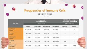 挂图Frequencies of Immune Cells in Rat Tissue Lists the estimated frequencies of more than 15 immune cell types in Sprague Dawley rats

 沪公网安备31010102008431号