Schreiber A et al. (JUL 2005)
Journal of the American Society of Nephrology : JASN 16 7 2216--24
Membrane proteinase 3 expression in patients with Wegener's granulomatosis and in human hematopoietic stem cell-derived neutrophils.
A large membrane proteinase 3 (mPR3)-positive neutrophil subset (mPR3high) is a risk for Wegener's granulomatosis (WG). The relationship between mPR3 expression and clinical manifestations was investigated in 81 WG patients and mPR3 expression was studied in CD34+ stem cell-derived human neutrophils. The mPR3high neutrophil percentage correlated with renal function,anemia,and albumin at the time of presentation. The mPR3high neutrophil percentage and renal failure severity correlated directly after 5 yr. For elucidating mechanisms that govern mPR3 expression,studies were conducted to determine whether the genetic information that governs mPR3 expression resides within the neutrophils,even without stimuli possibly related to disease. CD34+ hematopoietic stem cells were differentiated to neutrophils,and their mPR3 expression was determined. A two-step amplification/differentiation protocol was used to differentiate human CD34+ hematopoietic stem cells into neutrophils with G-CSF. The cells progressively expressed the neutrophil surface markers CD66b,CD35,and CD11b. The ferricytochrome C assay demonstrated a strong respiratory burst at day 14 in response to PMA but none at day 0. Intracellular PR3 was detectable from day 4 by Western blotting. An increasing percentage of a mPR3-positive neutrophil subset became detectable by flow cytometry,whereas a second subset remained negative,consistent with a bimodal expression. Finally,human PR3-anti-neutrophil cytoplasmic autoantibodies induced a stronger respiratory burst,compared with human control IgG in stem cell-derived neutrophils. Taken together,these studies underscore the clinical importance of the WG mPR3 phenotype. The surface mPR3 on resting cells is probably genetically determined rather than being dictated by external factors.
View Publication
Maes C et al. (MAY 2006)
The Journal of clinical investigation 116 5 1230--42
Placental growth factor mediates mesenchymal cell development, cartilage turnover, and bone remodeling during fracture repair.
Current therapies for delayed- or nonunion bone fractures are still largely ineffective. Previous studies indicated that the VEGF homolog placental growth factor (PlGF) has a more significant role in disease than in health. Therefore we investigated the role of PlGF in a model of semi-stabilized bone fracture healing. Fracture repair in mice lacking PlGF was impaired and characterized by a massive accumulation of cartilage in the callus,reminiscent of delayed- or nonunion fractures. PlGF was required for the early recruitment of inflammatory cells and the vascularization of the fracture wound. Interestingly,however,PlGF also played a role in the subsequent stages of the repair process. Indeed in vivo and in vitro findings indicated that PlGF induced the proliferation and osteogenic differentiation of mesenchymal progenitors and stimulated cartilage turnover by particular MMPs. Later in the process,PlGF was required for the remodeling of the newly formed bone by stimulating osteoclast differentiation. As PlGF expression was increased throughout the process of bone repair and all the important cell types involved expressed its receptor VEGFR-1,the present data suggest that PlGF is required for mediating and coordinating the key aspects of fracture repair. Therefore PlGF may potentially offer therapeutic advantages for fracture repair.
View Publication
产品号#:
03534
03334
03434
03444
18753
18753RF
产品名:
MethoCult™ GF M3534
MethoCult™ M3334
MethoCult™ GF M3434
MethoCult™ GF M3434
Kim M-H et al. (MAR 2011)
Blood 117 12 3343--52
Neutrophil survival and c-kit(+)-progenitor proliferation in Staphylococcus aureus-infected skin wounds promote resolution.
Polymorphonuclear neutrophils (PMNs) are critical for the formation,maintenance,and resolution of bacterial abscesses. However,the mechanisms that regulate PMN survival and proliferation during the evolution of an abscess are not well defined. Using a mouse model of Staphylococcus aureus abscess formation within a cutaneous wound,combined with real-time imaging of genetically tagged PMNs,we observed that a high bacterial burden elicited a sustained mobilization of PMNs from the bone marrow to the infected wound,where their lifespan was markedly extended. A continuous rise in wound PMN number,which was not accounted for by trafficking from the bone marrow or by prolonged survival,was correlated with the homing of c-kit(+)-progenitor cells from the blood to the wound,where they proliferated and formed mature PMNs. Furthermore,by blocking their recruitment with an antibody to c-kit,which severely limited the proliferation of mature PMNs in the wound and shortened mouse survival,we confirmed that progenitor cells are not only important contributors to PMN expansion in the wound,but are also functionally important for immune protection. We conclude that the abscess environment provides a niche capable of regulating PMN survival and local proliferation of bone marrow-derived c-kit(+)-progenitor cells.
View Publication
产品号#:
03434
03444
产品名:
MethoCult™ GF M3434
MethoCult™ GF M3434
Elling C et al. (MAR 2011)
Blood 117 10 2935--43
Novel imatinib-sensitive PDGFRA-activating point mutations in hypereosinophilic syndrome induce growth factor independence and leukemia-like disease.
The FIP1L1-PDGFRA fusion is seen in a fraction of cases with a presumptive diagnosis of hypereosinophilic syndrome (HES). However,because most HES patients lack FIP1L1-PDGFRA,we studied whether they harbor activating mutations of the PDGFRA gene. Sequencing of 87 FIP1L1-PDGFRA-negative HES patients revealed several novel PDGFRA point mutations (R481G,L507P,I562M,H570R,H650Q,N659S,L705P,R748G,and Y849S). When cloned into 32D cells,N659S and Y849S and-on selection for high expressors-also H650Q and R748G mutants induced growth factor-independent proliferation,clonogenic growth,and constitutive phosphorylation of PDGFRA and Stat5. Imatinib antagonized Stat5 phosphorylation. Mutations involving positions 659 and 849 had been shown previously to possess transforming potential in gastrointestinal stromal tumors. Because H650Q and R748G mutants possessed only weak transforming activity,we injected 32D cells harboring these mutants or FIP1L1-PDGFRA into mice and found that they induced a leukemia-like disease. Oral imatinib treatment significantly decreased leukemic growth in vivo and prolonged survival. In conclusion,our data provide evidence that imatinib-sensitive PDGFRA point mutations play an important role in the pathogenesis of HES and we propose that more research should be performed to further define the frequency and treatment response of PDGFRA mutations in FIP1L1-PDGFRA-negative HES patients.
View Publication
Deonarain R et al. (NOV 2003)
Proceedings of the National Academy of Sciences of the United States of America 100 23 13453--8
Critical roles for IFN-beta in lymphoid development, myelopoiesis, and tumor development: links to tumor necrosis factor alpha.
We have generated mice null for IFN-beta and report the diverse consequences of IFN-beta for both the innate and adaptive arms of immunity. Despite no abnormalities in the proportional balance of CD4 and CD8 T cell populations in the peripheral blood,thymus,and spleen of IFN-beta-/- mice,activated lymph node and splenic T lymphocytes exhibit enhanced T cell proliferation and decreased tumor necrosis factor alpha production,relative to IFN-beta+/+ mice. Notably,constitutive and induced expression of tumor necrosis factor alpha is reduced in the spleen and bone marrow (BM) macrophages,respectively,of IFN-beta-/- mice. We also observe an altered splenic architecture in IFN-beta-/- mice and a reduction in resident macrophages. We identify a potential defect in B cell maturation in IFN-beta-/- mice,associated with a decrease in B220+ve/high/CD43-ve BM-derived cells and a reduction in BP-1,IgM,and CD23 expression. Circulating IgM-,Mac-1-,and Gr-1-positive cells are also substantially decreased in IFN-beta-/- mice. The decrease in the numbers of circulating macrophages and granulocytes likely reflects defective maturation of primitive BM hematopoiesis in mice,shown by the reduction of colony-forming units,granulocyte-macrophage. We proceeded to evaluate the in vivo growth of malignant cells in the IFN-beta-/- background and give evidence that Lewis lung carcinoma-specific tumor growth is more aggressive in IFN-beta-/- mice. Taken altogether,our data suggest that,in addition to the direct growth-inhibitory effects on tumor cells,IFN-beta is required during different stages of maturation in the development of the immune system.
View Publication
产品号#:
03434
03444
产品名:
MethoCult™ GF M3434
MethoCult™ GF M3434
Decot V et al. (JAN 2008)
Bio-medical materials and engineering 18 1 Suppl S19--26
Chimerism analysis following nonmyeloablative stem cell transplantation using a new cell subset separation method: Robosep.
Chimerism analysis has become an important tool to manage patients in the peri-transplant period of allogenic stem cell transplantation. During this period,cells of donor and host origin can coexist and increasing proportion of cells of host origin is considered as a recurrence of the underlying disease. We currently performed chimerism analysis on separate peripheral blood cell subsets,lymphocytes and granulocytes. To improve our isolation method,a new automated device from Stem Cell Technology Roboseptrade mark was tested and compared to our manual separation technique. The results obtained on T cell purification showed an improvement of the purity (98.42% with Robosep vs. 92.42% with the manual technique Rosettesep) and of the recovery (63.43% with Robosep and 38% with Rosettesep). The results were significantly improved on patient samples with less than 10% CD3 positive cells (purity: 90% vs. 44.44%; recovery: 73.79% vs. 43.98%). Granulocytes separation was based on CD15 expression. The results showed an improvement of the purity with Robosep (96.90% vs. 86.20% with the manual technique Polymorphprep) but the recovery was impaired (35.2% vs. 52.30%). Using a myeloid (CD66/CD33) cocktail,recovery was improved with the Robosep device (64.04% with the myeloid cocktail vs. 22.4% with the CD15 cocktail). Our data demonstrated that Robosep allowed a performant cell purification in the early period post-transplantation even for populations representing less than 10% of the peripheral blood cells.
View Publication
产品号#:
19051
19051RF
21000
20119
20155
18681
18681RF
产品名:
EasySep™人T细胞富集试剂盒
RoboSep™ 人T细胞富集试剂盒含滤芯吸头
RoboSep™- S
RoboSep™ 吸头组件抛光剂
RoboSep™分选管套装(9个塑料管)
von Vietinghoff S et al. (MAY 2007)
Blood 109 10 4487--93
NB1 mediates surface expression of the ANCA antigen proteinase 3 on human neutrophils.
Antineutrophil cytoplasmic antibodies (ANCAs) with specificity for proteinase 3 (PR3) are central to a form of ANCA-associated vasculitis. Membrane PR3 (mPR3) is expressed only on a subset of neutrophils. The aim of this study was to determine the mechanism of PR3 surface expression on human neutrophils. Neutrophils were isolated from patients and healthy controls,and hematopoietic stem cells from cord blood served as a model of neutrophil differentiation. Surface expression was analyzed by flow cytometry and confocal microscopy,and proteins were analyzed by Western blot experiments. Neutrophil subsets were separated by magnetic cell sorting. Transfection experiments were carried out in HEK293 and HL60 cell lines. Using neutrophils from healthy donors,patients with vasculitis,and neutrophilic differentiated stem cells we found that mPR3 display was restricted to cells expressing neutrophil glycoprotein NB1,a glycosylphosphatidylinositol (GPI)-linked surface receptor. mPR3 expression was decreased by enzymatic removal of GPI anchors from cell membranes and was absent in a patient with paroxysmal nocturnal hemoglobinuria. PR3 and NB1 coimmunoprecipitated from and colocalized on the neutrophil plasma membrane. Transfection with NB1 resulted in specific PR3 surface binding in different cell types. We conclude that PR3 membrane expression on neutrophils is mediated by the NB1 receptor.
View Publication
产品号#:
09600
09650
产品名:
StemSpan™ SFEM
StemSpan™ SFEM
Gibbs BF et al. (MAR 2008)
Clinical and experimental allergy : journal of the British Society for Allergy and Clinical Immunology 38 3 480--5
A rapid two-step procedure for the purification of human peripheral blood basophils to near homogeneity.
BACKGROUND: Basophils are increasingly utilized as indicators of allergic inflammation and as primary allergic effector cells to study signalling pathways. However,until the present,their enrichment has been time consuming,costly and limited to relatively few specialized laboratories. OBJECTIVE: We have therefore devised a reproducible and rapid method for the purification of human basophils from small quantities of peripheral blood within 1.5 h,which does not require the use of specialized equipment such as elutriators. METHODS: Human basophils were obtained from healthy volunteers undergoing venipuncture. Heparinized or K3-ethylenediaminetetraacetic acid blood samples were first subjected to centrifugation in Hetasep,directly followed by negative selection using immunomagnetic beads. Basophil morphology and purity were assessed by May-Grünwald staining of cytospins. IgE-mediated histamine release was analysed spectrofluorometrically and IL-4 and IL-13 production by quantitative RT-PCR. CD203c and CD63 surface expression was measured using flow cytometry before and after activation with anti-IgE. RESULTS: Using this protocol,basophils were enriched close to homogeneity in most cases with a mean purity of 99.34+/-0.88% (range 97-100%,n=18) and a mean recovery of 75.6 (range 39-100%,n=8). Basophil viability following purification was 99.6+/-0.89% using Trypan blue exclusion. The purification procedure gave rise to basophils with normal functional responses to anti-IgE regarding histamine release as well as IL-4 and IL-13 mRNA expression. Moreover,constitutive cell-surface CD203c/CD63 expressions were not elevated before anti-IgE stimulation. CONCLUSION: The rapidity,simplicity and reproducibility of this method will facilitate the employment of basophils in high-output ex vivo studies.
View Publication
产品号#:
19069
19069RF
产品名:
Kunishima S et al. (MAR 2008)
Blood 111 6 3015--23
Differential expression of wild-type and mutant NMMHC-IIA polypeptides in blood cells suggests cell-specific regulation mechanisms in MYH9 disorders.
MYH9 disorders such as May-Hegglin anomaly are characterized by macrothrombocytopenia and cytoplasmic granulocyte inclusion bodies that result from mutations in MYH9,the gene for nonmuscle myosin heavy chain-IIA (NMMHC-IIA). We examined the expression of mutant NMMHC-IIA polypeptide in peripheral blood cells from patients with MYH9 5770delG and 5818delG mutations. A specific antibody to mutant NMMHC-IIA (NT629) was raised against the abnormal carboxyl-terminal residues generated by 5818delG. NT629 reacted to recombinant 5818delG NMMHC-IIA but not to wild-type NMMHC-IIA,and did not recognize any cellular components of normal peripheral blood cells. Immunofluorescence and immunoblotting revealed that mutant NMMHC-IIA was present and sequestrated only in inclusion bodies within neutrophils,diffusely distributed throughout lymphocyte cytoplasm,sparsely localized on a diffuse cytoplasmic background in monocytes,and uniformly distributed at diminished levels only in large platelets. Mutant NMMHC-IIA did not translocate to lamellipodia in surface activated platelets. Wild-type NMMHC-IIA was homogeneously distributed among megakaryocytes derived from the peripheral blood CD34(+) cells of patients,but coarse mutant NMMHC-IIA was heterogeneously scattered without abnormal aggregates in the cytoplasm. We show the differential expression of mutant NMMHC-IIA and postulate that cell-specific regulation mechanisms function in MYH9 disorders.
View Publication
产品号#:
09600
09650
产品名:
StemSpan™ SFEM
StemSpan™ SFEM
Joulia R et al. (JAN 2015)
Nature communications 6 6174
Mast cells form antibody-dependent degranulatory synapse for dedicated secretion and defence.
Mast cells are tissue-resident immune cells that play a key role in inflammation and allergy. Here we show that interaction of mast cells with antibody-targeted cells induces the polarized exocytosis of their granules resulting in a sustained exposure of effector enzymes,such as tryptase and chymase,at the cell-cell contact site. This previously unidentified mast cell effector mechanism,which we name the antibody-dependent degranulatory synapse (ADDS),is triggered by both IgE- and IgG-targeted cells. ADDSs take place within an area of cortical actin cytoskeleton clearance in the absence of microtubule organizing centre and Golgi apparatus repositioning towards the stimulating cell. Remarkably,IgG-mediated degranulatory synapses also occur upon contact with opsonized Toxoplasma gondii tachyzoites resulting in tryptase-dependent parasite death. Our results broaden current views of mast cell degranulation by revealing that human mast cells form degranulatory synapses with antibody-targeted cells and pathogens for dedicated secretion and defence.
View Publication

 EasySep™小鼠TIL(CD45)正选试剂盒


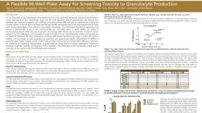 科学海报A Flexible 96-Well Plate Assay for Screening Toxicity to Granulocyte Production

 沪公网安备31010102008431号