D. R. McHugh et al. ( 2018)
PloS one 13 6 e0199573
A G542X cystic fibrosis mouse model for examining nonsense mutation directed therapies.
Nonsense mutations are present in 10{\%} of patients with CF,produce a premature termination codon in CFTR mRNA causing early termination of translation,and lead to lack of CFTR function. There are no currently available animal models which contain a nonsense mutation in the endogenous Cftr locus that can be utilized to test nonsense mutation therapies. In this study,we create a CF mouse model carrying the G542X nonsense mutation in Cftr using CRISPR/Cas9 gene editing. The G542X mouse model has reduced Cftr mRNA levels,demonstrates absence of CFTR function,and displays characteristic manifestations of CF mice such as reduced growth and intestinal obstruction. Importantly,CFTR restoration is observed in G542X intestinal organoids treated with G418,an aminoglycoside with translational readthrough capabilities. The G542X mouse model provides an invaluable resource for the identification of potential therapies of CF nonsense mutations as well as the assessment of in vivo effectiveness of these potential therapies targeting nonsense mutations.
View Publication
产品号#:
06005
产品名:
IntestiCult™ 类器官生长培养基 (小鼠)
Y. Sei et al. (MAY 2018)
American journal of physiology. Gastrointestinal and liver physiology
Mature Enteroendocrine Cells Contributes to Basal and Pathological Stem Cell Dynamics in the Small Intestine.
Lgr5-expressing intestinal stem cells (ISCs) maintain continuous and rapid generation of the intestinal epithelium. Here we present evidence that dedifferentiation of committed enteroendocrine cells (EECs) contributes to maintenance of the epithelium under both basal conditions and in response to injury. Lineage tracing studies identified a subset of EECs that reside at +4 position for more than 2 weeks,most of which were BrdU-label-retaining cells. Under basal conditions,cells derived from these EECs grow from the bottom of the crypt to generate intestinal epithelium according to neutral drift kinetics that is consistent with dedifferentiation of mature EECs to ISCs. The lineage tracing of EECs demonstrated reserve stem cell properties in response to radiation-induced injury with the generation of reparative EEC-derived epithelial patches. Finally,the enterochromaffin (EC) cell was the predominant EEC type participating in these stem cell dynamics. These results provide novel insights into the +4 reserve ISC hypothesis,stem cell dynamics of the intestinal epithelium and novel insight in the development of EC-derived small intestinal tumors.
View Publication
产品号#:
06005
产品名:
IntestiCult™ 类器官生长培养基 (小鼠)
D. Sharma et al. (DEC 2018)
Gastroenterology 154 4 948--964.e8
Pyrin Inflammasome Regulates Tight Junction Integrity to Restrict Colitis and Tumorigenesis.
BACKGROUND & AIMS Inflammatory bowel diseases (IBD) increase risk for colorectal cancer. Mutations in the Mediterranean fever gene (MEFV or pyrin) are associated with hereditary autoinflammatory disease and severe IBD. Expression of MEFV,a sensor protein that the initiates assembly of the inflammasome complex,is increased in colon biopsies from patients with IBD. We investigated the role of pyrin in intestinal homeostasis in mice. METHODS Mefv-/- mice and C57/BL6 mice (controls) were given azoxymethane followed by multiple rounds of dextran sodium sulfate (DSS) to induce colitis and tumorigenesis. In some experiments,Mefv-/- mice were given injections of recombinant interleukin 18 (rIL18) or saline (control) during DSS administration. Colon tissues were collected at different time points during colitis development and analyzed by histology,immunohistochemistry,immunoblots,or ELISAs (to measure cytokines). Spleen and mesenteric lymph node were collected,processed,and analyzed by flow cytometry. Colon epithelial permeability was measured in mice with colitis by gavage of fluorescent dextran and quantification of serum levels. RESULTS MEFV was expressed in colons of control mice and expression increased during chronic and acute inflammation; high levels were detected in colon tumor and adjacent non-tumor tissues. Mefv-/- mice developed more severe colitis than control mice,with a greater extent of epithelial hyperplasia and a larger tumor burden. Levels of inflammatory cytokines (IL6) and chemokines were significantly higher in colons of Mefv-/- mice than control mice following colitis induction,whereas the level IL18,which depends on the inflammasome for maturation and release,was significantly lower in colons of Mefv-/- mice. Mefv-/- mice had increased epithelial permeability following administration of DSS than control mice,and loss of the tight junction proteins occludin and claudin-2 from intercellular junctions. STAT3 was activated (phosphorylated) in inflamed colon tissues from Mefv-/-,which also had increased expression of stem cell markers (OLFM4,BMI1,and MSI1) compared with colons from control mice. Administration of rIL18 to Mefv-/- mice reduced epithelial permeability,intestinal inflammation,the severity of colitis,and colon tumorigenesis. CONCLUSIONS In studies with DSS-induced colitis,we found that pyrin (MEFV) is required for inflammasome activation and IL18 maturation,which promote intestinal barrier integrity and prevent colon inflammation and tumorigenesis. Strategies to increase activity of MEFV or IL18 might be developed for the treatment of IBD and prevention of colitis-associated tumorigenesis.
View Publication
产品号#:
06005
产品名:
IntestiCult™ 类器官生长培养基 (小鼠)
Tomé et al. (AUG 2016)
The Journal of nutritional biochemistry 34 146--55
Hydroxytyrosol supplementation modulates the expression of miRNAs in rodents and in humans.
Dietary microRNAs (miRNAs) modulation could be important for health and wellbeing. Part of the healthful activities of polyphenols might be due to a modulation of miRNAs' expression. Among the most biologically active polyphenols,hydroxytyrosol (HT) has never been studied for its actions on miRNAs. We investigated whether HT could modulate the expression of miRNAs in vivo. We performed an unbiased intestinal miRNA screening in mice supplemented (for 8 weeks) with nutritionally relevant amounts of HT. HT modulated the expression of several miRNAs. Analysis of other tissues revealed consistent HT-induced modulation of only few miRNAs. Also,HT administration increased triglycerides levels. Acute treatment with HT and in vitro experiments provided mechanistic insights. The HT-induced expression of one miRNA was confirmed in healthy volunteers supplemented with HT in a randomized,double-blind and placebo-controlled trial. HT consumption affects specific miRNAs' expression in rodents and humans. Our findings suggest that the modulation of miRNAs' action through HT consumption might partially explain its healthful activities and might be pharmanutritionally exploited in current therapies targeting endogenous miRNAs. However,the effects of HT on triglycerides warrant further investigations.
View Publication
Increased Abundance of M Cells in the Gut Epithelium Dramatically Enhances Oral Prion Disease Susceptibility.
Many natural prion diseases of humans and animals are considered to be acquired through oral consumption of contaminated food or pasture. Determining the route by which prions establish host infection will identify the important factors that influence oral prion disease susceptibility and to which intervention strategies can be developed. After exposure,the early accumulation and replication of prions within small intestinal Peyer's patches is essential for the efficient spread of disease to the brain. To replicate within Peyer's patches,the prions must first cross the gut epithelium. M cells are specialised epithelial cells within the epithelia covering Peyer's patches that transcytose particulate antigens and microorganisms. M cell-development is dependent upon RANKL-RANK-signalling,and mice in which RANK is deleted only in the gut epithelium completely lack M cells. In the specific absence of M cells in these mice,the accumulation of prions within Peyer's patches and the spread of disease to the brain was blocked,demonstrating a critical role for M cells in the initial transfer of prions across the gut epithelium in order to establish host infection. Since pathogens,inflammatory stimuli and aging can modify M cell-density in the gut,these factors may also influence oral prion disease susceptibility. Mice were therefore treated with RANKL to enhance M cell density in the gut. We show that prion uptake from the gut lumen was enhanced in RANKL-treated mice,resulting in shortened survival times and increased disease susceptibility,equivalent to a 10-fold higher infectious titre of prions. Together these data demonstrate that M cells are the critical gatekeepers of oral prion infection,whose density in the gut epithelium directly limits or enhances disease susceptibility. Our data suggest that factors which alter M cell-density in the gut epithelium may be important risk factors which influence host susceptibility to orally acquired prion diseases.
View Publication
产品号#:
06005
产品名:
IntestiCult™ 类器官生长培养基 (小鼠)
Hahn S et al. (MAY 2017)
Scientific reports 7 1 2435
Organoid-based epithelial to mesenchymal transition (OEMT) model: from an intestinal fibrosis perspective.
The current in vitro or in vivo intestinal fibrosis models have many limitations. Recent advancements in the isolation and culturing of organoids has led to development of various three-dimensional (3D) intestinal disease models with in vivo physiology. In this study,we generated an organoid-based epithelial to mesenchymal transition (OEMT) model,which could be used as a novel intestinal fibrosis model. Intestinal epithelial organoids (IEOs) were isolated and cultured from the small intestines of normal mice. IEOs were treated with transforming growth factor- β1 (TGF-β1) or Tumor necrosis factor-α (TNF-α) to evaluate their phenotypic change. Raw 264.7 cells (macrophage) stimulated with lipopolysaccharide were co-cultured with IEOs in growth media with or without TGF-β1. TGF-β1 alone slightly induced epithelial to mesenchymal transition (EMT) in the IEOs but mainly disrupted them. Macrophage released cytokines synergistically induced mesenchymal phenotypic changes in TGF-β1 stimulated intestinal organoids. TNF-α and TGF-β1 synergistically induced proliferation of mesenchymal cells as well as EMT in the IEOs. We generated a novel OEMT model based on our finding that TNF-α and TGF-β synergistically induce type 2 EMT in IEOs. This 3D EMT model with in vivo physiology could be used to study EMT associated intestinal fibrosis.
View Publication
产品号#:
06005
产品名:
IntestiCult™ 类器官生长培养基 (小鼠)
McIntyre BAS et al. (JUL 2015)
Innate immunity 21 5 504--511
Innate immune response of human pluripotent stem cell-derived airway epithelium.
The acquisition of innate immune response is requisite to having bona fide differentiation of airway epithelium. Procedures developed to differentiate lung airway from human pluripotent stem cells (hPSCs) have demonstrated anecdotal evidence for innate immune response,but an in-depth exploration of response levels is lacking. Herein,using an established method of airway epithelial generation from hPSCs,we show that hPSC-derived epithelial cells are able to up-regulate expression of TNF$\$,IL8 and IL1$\$ response to challenge with bacterial endotoxin LPS,but lack response from genes associated with innate immune response in other cell types. Further,stimulation of cells with TNF-$\$ in auto-induction of TNF$\$,as well as cytokine responses of IL8 and IL1$\$ The demonstration of innate immune induction in hPSC-derived airway epithelia gives further strength to the functionality of in vitro protocols aimed at generating differentiated airway cells that can potentially be used in a translational setting. Finally,we propose that innate immune challenge of airway epithelium from human pluripotent stem cell sources be used as a robust validation of functional in vitro differentiation.
View Publication
产品号#:
05850
05857
05870
05875
85850
85857
85870
85875
产品名:
mTeSR™1
mTeSR™1
Hansson ML et al. (FEB 2015)
Journal of Biological Chemistry 290 9 5661--5672
Efficient delivery and functional expression of transfected modified mRNA in human embryonic stem cell-derived retinal pigmented epithelial cells
Gene- and cell-based therapies are promising strategies for the treatment of degenerative retinal diseases such as age-related macular degeneration,Stargardt disease,and retinitis pigmentosa. Cellular engineering before transplantation may allow the delivery of cellular factors that can promote functional improvements,such as increased engraftment or survival of transplanted cells. A current challenge in traditional DNA-based vector transfection is to find a delivery system that is both safe and efficient,but using mRNA as an alternative to DNA can circumvent these major roadblocks. In this study,we show that both unmodified and modified mRNA can be delivered to retinal pigmented epithelial (RPE) cells with a high efficiency compared with conventional plasmid delivery systems. On the other hand,administration of unmodified mRNA induced a strong innate immune response that was almost absent when using modified mRNA. Importantly,transfection of mRNA encoding a key regulator of RPE gene expression,microphthalmia-associated transcription factor (MITF),confirmed the functionality of the delivered mRNA. Immunostaining showed that transfection with either type of mRNA led to the expression of roughly equal levels of MITF,primarily localized in the nucleus. Despite these findings,quantitative RT-PCR analyses showed that the activation of the expression of MITF target genes was higher following transfection with modified mRNA compared with unmodified mRNA. Our findings,therefore,show that modified mRNA transfection can be applied to human embryonic stem cell-derived RPE cells and that the method is safe,efficient,and functional.
View Publication
产品号#:
05850
05857
05870
05875
85850
85857
85870
85875
产品名:
mTeSR™1
mTeSR™1
Kumar A et al. (JAN 2012)
Breast cancer research : BCR 14 1 R4
Evidence that GTP-binding domain but not catalytic domain of transglutaminase 2 is essential for epithelial-to-mesenchymal transition in mammary epithelial cells.
INTRODUCTION: The expression of proinflammatory protein tissue transglutaminase 2 (TG2) is frequently upregulated in multiple cancer cell types. However,the exact role of TG2 in cancer cells is not well-understood. We recently initiated studies to determine the significance of TG2 in cancer cells and observed that sustained expression of TG2 resulted in epithelial-to-mesenchymal transition (EMT) and promoted cancer stem cell (CSC) traits in mammary epithelial cells. These results suggested that TG2 could serve as a promising therapeutic target for overcoming chemoresistance and inhibiting metastatic spread of cancer cells. METHODS: Using various mutant constructs,we analyzed the activity of TG2 that is essential for promoting the EMT-CSC phenotype. RESULTS: Our results suggest that catalytically inactive TG2 (TG2-C277S) is as effective as wild-type TG2 (TG2-WT) in inducing the EMT-CSC in mammary epithelial cells. In contrast,overexpression of a GTP-binding-deficient mutant (TG2-R580A) was completely incompetent in this regard. Moreover,TG2-dependent activation of the proinflammatory transcription factor NF-κB is deemed essential for promoting the EMT-CSC phenotype in mammary epithelial cells. CONCLUSIONS: Our results suggest that the transamidation activity of TG2 is not essential for promoting its oncogenic functions and provide a strong rationale for developing small-molecule inhibitors to block GTP-binding pockets of TG2. Such inhibitors may have great potential for inhibiting the TG2-regulated pathways,reversing drug resistance and inhibiting the metastasis of cancer cells.
View Publication

 EasySep™小鼠TIL(CD45)正选试剂盒


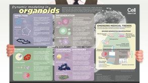 挂图Dynamic Modeling in Organoids Learn more about organoid applications for studying human health

 沪公网安备31010102008431号