
 EasySep™小鼠TIL(CD45)正选试剂盒


技术资料
-
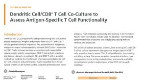 技术公告Dendritic Cell/CD8+ T Cell Co-Culture to Assess Antigen-Specific T Cell Functionality
细胞类型:
CD8+ T细胞,T细胞,单核细胞,树突状细胞(DCs)
发布日期: 11/22/2023 -
 37:09
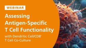
线上讲座Quick and Easy Isolation of T Cells and Other Immune Cells from Large-Volume Samples发布日期: 05/27/2022
37:09
线上讲座Quick and Easy Isolation of T Cells and Other Immune Cells from Large-Volume Samples发布日期: 05/27/2022 -


 沪公网安备31010102008431号