
 EasySep™小鼠TIL(CD45)正选试剂盒


技术资料
-
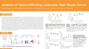 科学海报Isolation of Tumor-Infiltrating Leukocytes from Mouse Tumors
Conference:
AAI 2020
发布日期: 10/22/2020 -
-

-

-

-

-

若您需要咨询产品或有任何技术问题,请通过官方电话 400 885 9050 或邮箱 info.cn@stemcell.com 与我们联系。
Conference:
AAI 2020
在线联系


 沪公网安备31010102008431号