Carrera Silva EA et al. ( 2017)
Blood 130 17 1898--1902
CD207+CD1a+ cells circulate in pediatric patients with active Langerhans cell histiocytosis.
Langerhans cell histiocytosis (LCH) is a rare disease with an unknown etiology characterized by heterogeneous lesions containing CD207+CD1a+ cells that can arise in almost any tissue and cause significant morbidity and mortality. Precursors of pathological Langerhans cells have yet to be defined. Our aim was to identify circulating CD207+CD1a+ cells and their inducers in LCH. Expression of CD207 and CD1a in the blood myeloid compartment as well as thymic stromal lymphopoietin (TSLP) and transforming growth factor β (TGF-β) plasma levels were measured in 22 pediatric patients with active disease (AD) or nonactive disease (NAD). In patients with AD vs those with NAD,the myeloid compartment showed an increased CD11b (CD11bhigh plus CD11b+) fraction (39.7 ± 3.6 vs 18.6 ± 1.9),a higher percentage of circulating CD11bhighCD11c+CD207+ cells (44.5 ± 11.3 vs 3.2 ± 0.5),and the presence of CD11chighCD207+CD1a+ cells (25.0 ± 9.1 vs 2.3 ± 0.5). Blood CD207+CD1a+ cells were not observed in adult controls or umbilical cord. Increased TSLP and TGF-β levels were detected in patients with AD. Interestingly,plasma from patients with AD induces CD207 expression on CD14+ monocytes. We conclude that CD207+CD1a+ cells are circulating in patients with active LCH,and TSLP and TGF-β are potential drivers of Langerhans-like cells in vivo.
View Publication
产品号#:
17858
17858RF
100-0694
产品名:
EasySep™人CD14正选试剂盒II
RoboSep™ 人CD14正选试剂盒II
EasySep™人CD14正选试剂盒II
Kovats S et al. (NOV 2016)
Clinical and experimental immunology 186 2 214--226
West Nile virus-infected human dendritic cells fail to fully activate invariant natural killer T cells.
West Nile virus (WNV) infection is a mosquito-borne zoonosis with increasing prevalence in the United States. WNV infection begins in the skin,and the virus replicates initially in keratinocytes and dendritic cells (DCs). In the skin and cutaneous lymph nodes,infected DCs are likely to interact with invariant natural killer T cells (iNKTs). Bidirectional interactions between DCs and iNKTs amplify the innate immune response to viral infections,thus controlling viral load and regulating adaptive immunity. iNKTs are stimulated by CD1d-bound lipid antigens or activated indirectly by inflammatory cytokines. We exposed human monocyte-derived DCs to WNV Kunjin and determined their ability to activate isolated blood iNKTs. DCs became infected as judged by synthesis of viral mRNA and Envelope and NS-1 proteins,but did not undergo significant apoptosis. Infected DCs up-regulated the co-stimulatory molecules CD86 and CD40,but showed decreased expression of CD1d. WNV infection induced DC secretion of type I interferon (IFN),but no or minimal interleukin (IL)-12,IL-23,IL-18 or IL-10. Unexpectedly,we found that the WNV-infected DCs stimulated human iNKTs to up-regulate CD69 and produce low amounts of IL-10,but not proinflammatory cytokines such as IFN-γ or tumour necrosis factor (TNF)-α. Both CD1d and IFNAR blockade partially abrogated this iNKT response,suggesting involvement of a T cell receptor (TCR)-CD1d interaction and type I interferon receptor (IFNAR) signalling. Thus,WNV infection interferes with DC-iNKT interactions by preventing the production of proinflammatory cytokines. iNKTs may be a source of IL-10 observed in human flavivirus infections and initiate an anti-inflammatory innate response that limits adaptive immunity and immune pathology upon WNV infection.
View Publication
S100-alarmin-induced innate immune programming protects newborn infants from sepsis.
The high risk of neonatal death from sepsis is thought to result from impaired responses by innate immune cells; however,the clinical observation of hyperinflammatory courses of neonatal sepsis contradicts this concept. Using transcriptomic,epigenetic and immunological approaches,we demonstrated that high amounts of the perinatal alarmins S100A8 and S100A9 specifically altered MyD88-dependent proinflammatory gene programs. S100 programming prevented hyperinflammatory responses without impairing pathogen defense. TRIF-adaptor-dependent regulatory genes remained unaffected by perinatal S100 programming and responded strongly to lipopolysaccharide,but were barely expressed. Steady-state expression of TRIF-dependent genes increased only gradually during the first year of life in human neonates,shifting immune regulation toward the adult phenotype. Disruption of this critical sequence of transient alarmin programming and subsequent reprogramming of regulatory pathways increased the risk of hyperinflammation and sepsis. Collectively these data suggest that neonates are characterized by a selective,transient microbial unresponsiveness that prevents harmful hyperinflammation in the delicate neonate while allowing for sufficient immunological protection.
View Publication
Expression of specific inflammasome gene modules stratifies older individuals into two extreme clinical and immunological states.
Low-grade,chronic inflammation has been associated with many diseases of aging,but the mechanisms responsible for producing this inflammation remain unclear. Inflammasomes can drive chronic inflammation in the context of an infectious disease or cellular stress,and they trigger the maturation of interleukin-1β (IL-1β). Here we find that the expression of specific inflammasome gene modules stratifies older individuals into two extremes: those with constitutive expression of IL-1β,nucleotide metabolism dysfunction,elevated oxidative stress,high rates of hypertension and arterial stiffness; and those without constitutive expression of IL-1β,who lack these characteristics. Adenine and N(4)-acetylcytidine,nucleotide-derived metabolites that are detectable in the blood of the former group,prime and activate the NLRC4 inflammasome,induce the production of IL-1β,activate platelets and neutrophils and elevate blood pressure in mice. In individuals over 85 years of age,the elevated expression of inflammasome gene modules was associated with all-cause mortality. Thus,targeting inflammasome components may ameliorate chronic inflammation and various other age-associated conditions.
View Publication
产品号#:
15028
15068
产品名:
RosetteSep™人单核细胞富集抗体混合物
RosetteSep™人单核细胞富集抗体混合物
Li Q et al. (AUG 2005)
Proceedings of the National Academy of Sciences of the United States of America 102 35 12425--30
Enhanced NF-kappaB activation and cellular function in macrophages lacking IkappaB kinase 1 (IKK1).
IkappaB kinase (IKK) complex plays a key regulatory role in macrophages for NF-kappaB activation during both innate and adaptive immune responses. Because IKK1-/- mice died at birth,we differentiated functional macrophages from embryonic day 15.5 IKK1 mutant embryonic liver. The embryonic liver-derived macrophage (ELDM) showed enhanced phagocytotic clearance of bacteria,more efficient antigen-presenting capacity,elevated secretion of several key proinflammatory cytokines and chemokines,and known NFkappaB target genes. Increased NFkappaB activity in IKK1 mutant ELDM was the result of prolonged degradation of IkappaBalpha in response to infectious pathogens. The delayed restoration of IkappaBalpha in pathogen-activated IKK1-/- ELDM was a direct consequence of uncontrolled IKK2 kinase activity. We hypothesize that IKK1 plays a checkpoint role in the proper control of IkappaBalpha kinase activity in innate and adaptive immunity.
View Publication

 EasySep™小鼠TIL(CD45)正选试剂盒


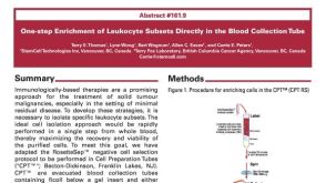 科学海报One-Step Enrichment of Leukocyte Subsets Directly in the Blood Collection Tube


 沪公网安备31010102008431号