Fuerstenau-Sharp M et al. (MAY 2015)
PloS one 10 5 e0126596
Generation of highly purified human cardiomyocytes from peripheral blood mononuclear cell-derived induced pluripotent stem cells.
Induced pluripotent stem (iPS) cells have an enormous potential for physiological studies. A novel protocol was developed combining the derivation of iPS from peripheral blood with an optimized directed differentiation to cardiomyocytes and a subsequent metabolic selection. The human iPS cells were retrovirally dedifferentiated from activated T cells. The subsequent optimized directed differentiation protocol yielded 30-45% cardiomyocytes at day 16 of differentiation. The derived cardiomyocytes expressed appropriate structural markers like cardiac troponin T,$\$-actinin and myosin light chain 2 (MLC2V). In a subsequent metabolic selection with lactate,the cardiomyocytes content could be increased to more than 90%. Loss of cardiomyocytes during metabolic selection were less than 50%,whereas alternative surface antibody-based selection procedures resulted in loss of up to 80% of cardiomyocytes. Electrophysiological characterization confirmed the typical cardiac features and the presence of ventricular,atrial and nodal-like action potentials within the derived cardiomyocyte population. Our combined and optimized protocol is highly robust and applicable for scalable cardiac differentiation. It provides a simple and cost-efficient method without expensive equipment for generating large numbers of highly purified,functional cardiomyocytes. It will further enhance the applicability of iPS cell-derived cardiomyocytes for disease modeling,drug discovery,and regenerative medicine.
View Publication
产品号#:
05850
05857
05870
05875
85850
85857
85870
85875
产品名:
mTeSR™1
mTeSR™1
Iqbal AJ et al. (OCT 2014)
Blood 124 15 e33--44
Human CD68 promoter GFP transgenic mice allow analysis of monocyte to macrophage differentiation in vivo.
The recruitment of monocytes and their differentiation into macrophages at sites of inflammation are key events in determining the outcome of the inflammatory response and initiating the return to tissue homeostasis. To study monocyte trafficking and macrophage differentiation in vivo,we have generated a novel transgenic reporter mouse expressing a green fluorescent protein (GFP) under the control of the human CD68 promoter. CD68-GFP mice express high levels of GFP in both monocyte and embryo-derived tissue resident macrophages in adult animals. The human CD68 promoter drives GFP expression in all CD115(+) monocytes of adult blood,spleen,and bone marrow; we took advantage of this to directly compare the trafficking of bone marrow-derived CD68-GFP monocytes to that of CX3CR1(GFP) monocytes in vivo using a sterile zymosan peritonitis model. Unlike CX3CR1(GFP) monocytes,which downregulate GFP expression on differentiation into macrophages in this model,CD68-GFP monocytes retain high-level GFP expression for 72 hours after differentiation into macrophages,allowing continued cell tracking during resolution of inflammation. In summary,this novel CD68-GFP transgenic reporter mouse line represents a powerful resource for analyzing monocyte mobilization and monocyte trafficking as well as studying the fate of recruited monocytes in models of acute and chronic inflammation.
View Publication
产品号#:
18102
19761
19761RF
产品名:
EasyPlate™ EasySep™磁极
挂图
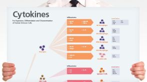
Human Immune Cytokines
Infographic of key cytokines for expansion, differentiation and characterization of major immune cell types
Liu Y-S et al. (MAY 2017)
Oncogene
MiR-181b modulates EGFR-dependent VCAM-1 expression and monocyte adhesion in glioblastoma.
Tumor-associated macrophages (TAMs) originate as circulating monocytes,and are recruited to gliomas,where they facilitate tumor growth and migration. Understanding the interaction between TAM and cancer cells may identify therapeutic targets for glioblastoma multiforme (GBM). Vascular cell adhesion molecule-1 (VCAM-1) is a cytokine-induced adhesion molecule expressed on the surface of cancer cells,which is involved in interactions with immune cells. Analysis of the glioma patient database and tissue immunohistochemistry showed that VCAM-1 expression correlated with the clinico-pathological grade of gliomas. Here,we found that VCAM-1 expression correlated positively with monocyte adhesion to GBM,and knockdown of VCAM-1 abolished the enhancement of monocyte adhesion. Importantly,upregulation of VCAM-1 is dependent on epidermal-growth-factor-receptor (EGFR) expression,and inhibition of EGFR effectively reduced VCAM-1 expression and monocyte adhesion activity. Moreover,GBM possessing higher EGFR levels (U251 cells) had higher VCAM-1 levels compared to GBMs with lower levels of EGFR (GL261 cells). Using two- and three-dimensional cultures,we found that monocyte adhesion to GBM occurs via integrin α4β1,which promotes tumor growth and invasion activity. Increased proliferation and tumor necrosis factor-α and IFN-γ levels were also observed in the adherent monocytes. Using a genetic modification approach,we demonstrated that VCAM-1 expression and monocyte adhesion were regulated by the miR-181 family,and lower levels of miR-181b correlated with high-grade glioma patients. Our results also demonstrated that miR-181b/protein phosphatase 2A-modulated SP-1 de-phosphorylation,which mediated the EGFR-dependent VCAM-1 expression and monocyte adhesion to GBM. We also found that the EGFR-dependent VCAM-1 expression is mediated by the p38/STAT3 signaling pathway. Our study suggested that VCAM-1 is a critical modulator of EGFR-dependent interaction of monocytes with GBM,which raises the possibility of developing effective and improved therapies for GBM.Oncogene advance online publication,1 May 2017; doi:10.1038/onc.2017.129.
View Publication
产品号#:
15028
15068
产品名:
RosetteSep™人单核细胞富集抗体混合物
RosetteSep™人单核细胞富集抗体混合物
Pereira RC et al. ( 2016)
Frontiers in immunology 7 415
Human Articular Chondrocytes Regulate Immune Response by Affecting Directly T Cell Proliferation and Indirectly Inhibiting Monocyte Differentiation to Professional Antigen-Presenting Cells.
Autologous chondrocyte implantation is the current gold standard cell therapy for cartilage lesions. However,in some instances,the heavily compromised health of the patient can either impair or limit the recovery of the autologous chondrocytes and a satisfactory outcome of the implant. Allogeneic human articular chondrocytes (hAC) could be a good alternative,but the possible immunological incompatibility between recipient and hAC donor should be considered. Herein,we report that allogeneic hAC inhibited T lymphocyte response to antigen-dependent and -independent proliferative stimuli. This effect was maximal when T cells and hAC were in contact and it was not relieved by the addition of exogenous lymphocyte growth factor interleukin (IL)-2. More important,hAC impaired the differentiation of peripheral blood monocytes induced with granulocyte monocyte colony-stimulating factor and IL-4 (Mo) to professional antigen-presenting cells,such as dendritic cells (DC). Indeed,a marked inhibition of the onset of the CD1a expression and an ineffective downregulation of CD14 antigens was observed in Mo-hAC co-cultures. Furthermore,compared to immature or mature DC,Mo from Mo-hAC co-cultures did not trigger an efficacious allo-response. The prostaglandin (PG) E2 present in the Mo-hAC co-culture conditioned media is a putative candidate of the hAC-mediated inhibition of Mo maturation. Altogether,these findings indicate that allogeneic hAC inhibit,rather than trigger,immune response and strongly suggest that an efficient chondrocyte implantation could be possible also in an allogeneic setting.
View Publication
产品号#:
17951
17951RF
17952
17952RF
18099
18099RF
100-0695
100-0696
产品名:
EasySep™人T细胞分选试剂盒
RoboSep™ 人T细胞分选试剂盒
EasySep™人CD4+ T细胞分选试剂盒
RoboSep™ 人CD4+ T细胞分选试剂盒
EasySep™人T细胞分选试剂盒
EasySep™人CD4+ T细胞分离试剂盒
Joseph J et al. ( 2016)
Nature communications 7 12748
Inhibition of ROS and upregulation of inflammatory cytokines by FoxO3a promotes survival against Salmonella typhimurium.
Virulent intracellular pathogens,such as the Salmonella species,engage numerous virulence factors to subvert host defence mechanisms to induce a chronic infection that leads to typhoid or exacerbation of other chronic inflammatory conditions. Here we show the role of the forkhead transcription factor FoxO3a during infection of mice with Salmonella typhimurium (ST). Although FoxO3a signalling does not affect the development of CD8(+) T cell responses to ST,FoxO3a has an important protective role,particularly during the chronic stage of infection,by limiting the persistence of oxidative stress. Furthermore,FoxO3a signalling regulates ERK signalling in macrophages,which results in the maintenance of a proinflammatory state. FoxO3a signalling does not affect cell proliferation or cell death. Thus,these results reveal mechanisms by which FoxO3a promotes host survival during infection with chronic,virulent intracellular bacteria.
View Publication
产品号#:
19761
19761RF
产品名:
Pourcet B et al. (MAY 2016)
Scientific Reports 6 25481
The nuclear receptor LXR modulates interleukin-18 levels in macrophages through multiple mechanisms.
IL-18 is a member of the IL-1 family involved in innate immunity and inflammation. Deregulated levels of IL-18 are involved in the pathogenesis of multiple disorders including inflammatory and metabolic diseases,yet relatively little is known regarding its regulation. Liver X receptors or LXRs are key modulators of macrophage cholesterol homeostasis and immune responses. Here we show that LXR ligands negatively regulate LPS-induced mRNA and protein expression of IL-18 in bone marrow-derived macrophages. Consistent with this being an LXR-mediated process,inhibition is abolished in the presence of a specific LXR antagonist and in LXR-deficient macrophages. Additionally,IL-18 processing of its precursor inactive form to its bioactive state is inhibited by LXR through negative regulation of both pro-caspase 1 expression and activation. Finally,LXR ligands further modulate IL-18 levels by inducing the expression of IL-18BP,a potent endogenous inhibitor of IL-18. This regulation occurs via the transcription factor IRF8,thus identifying IL-18BP as a novel LXR and IRF8 target gene. In conclusion,LXR activation inhibits IL-18 production through regulation of its transcription and maturation into an active pro-inflammatory cytokine. This novel regulation of IL-18 by LXR could be applied to modulate the severity of IL-18 driven metabolic and inflammatory disorders.
View Publication
Gilbert C et al. (JUL 2007)
Journal of virology 81 14 7672--82
Human immunodeficiency virus type 1 replication in dendritic cell-T-cell cocultures is increased upon incorporation of host LFA-1 due to higher levels of virus production in immature dendritic cells.
Dendritic cells (DCs) act as a portal for invasion by human immunodeficiency virus type-1 (HIV-1). Here,we investigated whether virion-incorporated host cell membrane proteins can affect virus replication in DC-T-cell cocultures. Using isogenic viruses either devoid of or bearing host-derived leukocyte function-associated antigen 1 (LFA-1),we showed that HIV-1 production is augmented when LFA-1-bearing virions are used compared to that for viral entities lacking this adhesion molecule. This phenomenon was observed in immature monocyte-derived DCs (IM-MDDCs) only and not in DCs displaying a mature phenotype. The increase is not due to higher virus production in responder CD4(+) T cells but rather is linked with a more important productive infection of IM-MDDCs. We provided evidence that virus-associated host LFA-1 molecules do not affect a late event in the HIV-1 life cycle but rather exert an effect on an early step in virus replication. We demonstrated that the enhancement of productive infection of IM-MDDCs that is conferred by virus-anchored host LFA-1 involves the protein kinase A (PKA) and PKC signal transduction pathways. The biological significance of this phenomenon was established by performing experiments with virus stocks produced in primary human cells and anti-LFA-1 antibodies. Together,our results indicate that the association between some virus-bound host proteins and their natural cognate ligands can modulate de novo HIV-1 production by IM-MDDCs. Therefore,the additional interactions between virus-bound host cell membrane constituents and counter receptors on the surfaces of DCs can influence HIV-1 replication in IM-MDDC-T-cell cocultures.
View Publication
产品号#:
18058
18058RF
19052
19052RF
产品名:
EasySep™人CD4+ T细胞富集试剂盒
RoboSep™ 人CD4+ T细胞富集试剂盒含滤芯吸头
Cai S et al. (NOV 2010)
Journal of immunology (Baltimore,Md. : 1950) 185 10 6214--25
CXCL1 regulates pulmonary host defense to Klebsiella Infection via CXCL2, CXCL5, NF-kappaB, and MAPKs.
Pulmonary bacterial infections are a leading cause of death. Since the introduction of antibiotics,multidrug-resistant Klebsiella pneumoniae became an escalating threat. Therefore,development of methods to augment antibacterial defense is warranted. Neutrophil recruitment is critical to clear bacteria,and neutrophil migration in the lung requires the production of ELR(+) CXC chemokines. Although lung-specific CXCL1/keratinocyte cell-derived chemokine (KC) transgene expression causes neutrophil-mediated clearance of K. pneumoniae,the mechanisms underlying KC-mediated host defense against K. pneumoniae have not been explored. In this study,we delineated the host defense functions of KC during pulmonary K. pneumoniae infection using KC(-/-) mice. Our findings demonstrate that KC is important for expression of CXCL2/MIP-2 and CXCL5/LPS-induced CXC chemokine,and activation of NF-κB and MAPKs in the lung. Furthermore,KC derived from both hematopoietic and resident cells contributes to host defense against K. pneumoniae. Neutrophil depletion in mice before K. pneumoniae infection reveals no differences in the production of MIP-2 and LPS-induced CXC chemokine or activation of NF-κB and MAPKs in the lung. Using murine bone marrow-derived and alveolar macrophages,we confirmed KC-mediated upregulation of MIP-2 and activation of NF-κB and MAPKs on K. pneumoniae infection. Moreover,neutralizing KC in bone marrow-derived macrophages before K. pneumoniae challenge decreases bacteria-induced production of KC and MIP-2,and activation of NF-κB and MAPKs. These findings reveal the importance of KC produced by hematopoietic and resident cells in regulating pulmonary host defense against a bacterial pathogen via the activation of transcription factors and MAPKs,as well as the expression of cell adhesion molecules and other neutrophil chemoattractants.
View Publication

 EasySep™小鼠TIL(CD45)正选试剂盒


 挂图Human Immune Cytokines Infographic of key cytokines for expansion, differentiation and characterization of major immune cell types
 实验方案How to Collect Plasma from Whole Blood Before Cell Isolation

 沪公网安备31010102008431号