Micropatterning Facilitates the Long-Term Growth and Analysis of iPSC-Derived Individual Human Neurons and Neuronal Networks
The discovery of induced pluripotent stem cells (iPSCs) and their application to patient-specific disease models offers new opportunities for studying the pathophysiology of neurological disorders. However,current methods for culturing iPSC-derived neuronal cells result in clustering of neurons,which precludes the analysis of individual neurons and defined neuronal networks. To address this challenge,cultures of human neurons on micropatterned surfaces are developed that promote neuronal survival over extended periods of time. This approach facilitates studies of neuronal development,cellular trafficking,and related mechanisms that require assessment of individual neurons and specific network connections. Importantly,micropatterns support the long-term stability of cultured neurons,which enables time-dependent analysis of cellular processes in living neurons. The approach described in this paper allows mechanistic studies of human neurons,both in terms of normal neuronal development and function,as well as time-dependent pathological processes,and provides a platform for testing of new therapeutics in neuropsychiatric disorders.
View Publication
产品号#:
05711
100-1281
产品名:
NeuroCult™ SM1 神经添加物
NeuroCult™ SM1 神经添加物
Ferreira JS et al. (JUN 2015)
The Journal of neuroscience : the official journal of the Society for Neuroscience 35 22 8462--79
GluN2B-Containing NMDA Receptors Regulate AMPA Receptor Traffic through Anchoring of the Synaptic Proteasome.
NMDA receptors play a central role in shaping the strength of synaptic connections throughout development and in mediating synaptic plasticity mechanisms that underlie some forms of learning and memory formation in the CNS. In the hippocampus and the neocortex,GluN1 is combined primarily with GluN2A and GluN2B,which are differentially expressed during development and confer distinct molecular and physiological properties to NMDA receptors. The contribution of each subunit to the synaptic traffic of NMDA receptors and therefore to their role during development and in synaptic plasticity is still controversial. We report a critical role for the GluN2B subunit in regulating NMDA receptor synaptic targeting. In the absence of GluN2B,the synaptic levels of AMPA receptors are increased and accompanied by decreased constitutive endocytosis of GluA1-AMPA receptor. We used quantitative proteomic analysis to identify changes in the composition of postsynaptic densities from GluN2B(-/-) mouse primary neuronal cultures and found altered levels of several ubiquitin proteasome system components,in particular decreased levels of proteasome subunits. Enhancing the proteasome activity with a novel proteasome activator restored the synaptic levels of AMPA receptors in GluN2B(-/-) neurons and their endocytosis,revealing that GluN2B-mediated anchoring of the synaptic proteasome is responsible for fine tuning AMPA receptor synaptic levels under basal conditions.
View Publication
产品号#:
05711
100-1281
产品名:
NeuroCult™ SM1 神经添加物
NeuroCult™ SM1 神经添加物
E. Hangen et al. (JUL 2018)
Cell reports 24 4 1001--1012.e3
Neuronal Activity and Intracellular Calcium Levels Regulate Intracellular Transport of Newly Synthesized AMPAR.
Regulation of AMPA receptor (AMPAR) trafficking is a key modulator of excitatory synaptic transmission; however,intracellular vesicular transport of newly synthesized AMPARs has been little studied due to technical limitations. By combining molecular tools with imaging strategies in cultured rat hippocampal neurons,we found that vesicles containing newly synthesized,GluA1-subunit-containing AMPARs are transported antero- and retrogradely at a mean speed of 1.5 mu$m.s-1. Synaptic activity and variations in intracellular calcium levels bidirectionally modulate GluA1 transport. Chemical long-term potentiation (cLTP) initially induces a halt in GluA1 transport,followed by a sustained increase,while acute glutamate uncaging on synaptic spines arrests vesicular movements. GluA1 phosphomimetic mutants preferentially travel to the dendritic tip,probably to replenish extrasynaptic pools,distal to the soma. Our findings indicate that AMPAR intracellular transport is highly regulated during synaptic plasticity and likely controls AMPAR numbers at the plasma membrane.
View Publication
A critical challenge to deciphering the pathophysiology of neurodevelopmental disease is identifying which of the myriad abnormalities that emerge during CNS maturation persist to contribute to long-term brain dysfunction. Childhood-onset dystonia caused by a loss-of-function mutation in the AAA+ protein torsinA exemplifies this challenge. Neurons lacking torsinA develop transient nuclear envelope (NE) malformations during CNS maturation,but no NE defects are described in mature torsinA null neurons. We find that during postnatal CNS maturation torsinA null neurons develop mislocalized and dysfunctional nuclear pore complexes (NPC) that lack NUP358,normally added late in NPC biogenesis. SUN1,a torsinA-related molecule implicated in interphase NPC biogenesis,also exhibits localization abnormalities. Whereas SUN1 and associated nuclear membrane abnormalities resolve in juvenile mice,NPC defects persist into adulthood. These findings support a role for torsinA function in NPC biogenesis during neuronal maturation and implicate altered NPC function in dystonia pathophysiology.
View Publication
产品号#:
05711
05790
05792
05793
05794
05795
100-1281
产品名:
NeuroCult™ SM1 神经添加物
BrainPhys™神经元培养基
BrainPhys™神经元培养基和SM1试剂盒
BrainPhys™ 神经元培养基N2-A和SM1试剂盒
BrainPhys™原代神经元试剂盒
BrainPhys™ hPSC 神经元试剂盒
NeuroCult™ SM1 神经添加物
Paquet D et al. (MAY 2016)
Nature 533 7601 125--129
Efficient introduction of specific homozygous and heterozygous mutations using CRISPR/Cas9
The bacterial CRISPR/Cas9 system allows sequence-specific gene editing in many organisms and holds promise as a tool to generate models of human diseases,for example,in human pluripotent stem cells. CRISPR/Cas9 introduces targeted double-stranded breaks (DSBs) with high efficiency,which are typically repaired by non-homologous end-joining (NHEJ) resulting in nonspecific insertions,deletions or other mutations (indels). DSBs may also be repaired by homology-directed repair (HDR) using a DNA repair template,such as an introduced single-stranded oligo DNA nucleotide (ssODN),allowing knock-in of specific mutations. Although CRISPR/Cas9 is used extensively to engineer gene knockouts through NHEJ,editing by HDR remains inefficient and can be corrupted by additional indels,preventing its widespread use for modelling genetic disorders through introducing disease-associated mutations. Furthermore,targeted mutational knock-in at single alleles to model diseases caused by heterozygous mutations has not been reported. Here we describe a CRISPR/Cas9-based genome-editing framework that allows selective introduction of mono- and bi-allelic sequence changes with high efficiency and accuracy. We show that HDR accuracy is increased dramatically by incorporating silent CRISPR/Cas-blocking mutations along with pathogenic mutations,and establish a method termed 'CORRECT' for scarless genome editing. By characterizing and exploiting a stereotyped inverse relationship between a mutation's incorporation rate and its distance to the DSB,we achieve predictable control of zygosity. Homozygous introduction requires a guide RNA targeting close to the intended mutation,whereas heterozygous introduction can be accomplished by distance-dependent suboptimal mutation incorporation or by use of mixed repair templates. Using this approach,we generated human induced pluripotent stem cells with heterozygous and homozygous dominant early onset Alzheimer's disease-causing mutations in amyloid precursor protein (APP(Swe)) and presenilin 1 (PSEN1(M146V)) and derived cortical neurons,which displayed genotype-dependent disease-associated phenotypes. Our findings enable efficient introduction of specific sequence changes with CRISPR/Cas9,facilitating study of human disease.
View Publication
产品号#:
05832
产品名:
STEMdiff™ 神经花环选择试剂
B. S. Souza et al. (dec 2016)
Scientific Reports 6 1 39775
Zika virus infection induces mitosis abnormalities and apoptotic cell death of human neural progenitor cells
Zika virus (ZIKV) infection has been associated with severe complications both in the developing and adult nervous system. To investigate the deleterious effects of ZIKV infection,we used human neural progenitor cells (NPC),derived from induced pluripotent stem cells (iPSC). We found that NPC are highly susceptible to ZIKV and the infection results in cell death. ZIKV infection led to a marked reduction in cell proliferation,ultrastructural alterations and induction of autophagy. Induction of apoptosis of Sox2 + cells was demonstrated by activation of caspases 3/7,8 and 9,and by ultrastructural and flow cytometry analyses. ZIKV-induced death of Sox2 + cells was prevented by incubation with the pan-caspase inhibitor,Z-VAD-FMK. By confocal microscopy analysis we found an increased number of cells with supernumerary centrosomes. Live imaging showed a significant increase in mitosis abnormalities,including multipolar spindle,chromosome laggards,micronuclei and death of progeny after cell division. FISH analysis for chromosomes 12 and 17 showed increased frequency of aneuploidy,such as monosomy,trisomy and polyploidy. Our study reinforces the link between ZIKV and abnormalities in the developing human brain,including microcephaly.
View Publication
产品号#:
05832
05833
19851
19851RF
19852
19852RF
19854
19854RF
05835
05839
产品名:
STEMdiff™ 神经花环选择试剂
STEMdiff™神经前体细胞培养基
EasySep™小鼠T细胞分选试剂盒
RoboSep™ 小鼠T细胞分选试剂盒
EasySep™小鼠CD4+ T细胞分选试剂盒
RoboSep™ 小鼠CD4+ T细胞分选试剂盒
EasySep™小鼠B细胞分选试剂盒
RoboSep™ 小鼠B细胞分选试剂盒
STEMdiff™ 神经诱导培养基
STEMdiff™ 神经诱导培养基
Tagliafierro L et al. (NOV 2017)
Alzheimer's & dementia : the journal of the Alzheimer's Association 13 11 1237--1250
Genetic analysis of α-synuclein 3' untranslated region and its corresponding microRNAs in relation to Parkinson's disease compared to dementia with Lewy bodies.
INTRODUCTION The α-synuclein (SNCA) gene has been implicated in the etiology of Parkinson's disease (PD) and dementia with Lewy bodies (DLB). METHODS A computational analysis of SNCA 3' untranslated region to identify potential microRNA (miRNA) binding sites and quantitative real-time polymerase chain reaction (PCR) to determine their expression in isogenic induced pluripotent stem cell-derived dopaminergic and cholinergic neurons as a model of PD and DLB,respectively,were performed. In addition,we performed a deep sequencing analysis of the SNCA 3' untranslated region of autopsy-confirmed cases of PD,DLB,and normal controls,followed by genetic association analysis of the identified variants. RESULTS We identified four miRNA binding sites and observed a neuronal-type-specific expression profile for each miRNA in the different isogenic induced pluripotent stem cell-derived dopaminergic and cholinergic neurons. Furthermore,we found that the short structural variant rs777296100-polyT was moderately associated with DLB but not with PD. DISCUSSION We suggest that the regulation of SNCA expression through miRNAs is neuronal-type-specific and possibly plays a part in the phenotypic heterogeneity of synucleinopathies. Furthermore,genetic variability in the SNCA gene may contribute to synucleinopathies in a pathology-specific manner.
View Publication
产品号#:
05790
05792
05793
05794
05795
产品名:
BrainPhys™神经元培养基
BrainPhys™神经元培养基和SM1试剂盒
BrainPhys™ 神经元培养基N2-A和SM1试剂盒
BrainPhys™原代神经元试剂盒
BrainPhys™ hPSC 神经元试剂盒
Werner A et al. (SEP 2015)
Nature 525 7570 523--527
Cell-fate determination by ubiquitin-dependent regulation of translation
Metazoan development depends on the accurate execution of differentiation programs that allow pluripotent stem cells to adopt specific fates. Differentiation requires changes to chromatin architecture and transcriptional networks,yet whether other regulatory events support cell-fate determination is less well understood. Here we identify the ubiquitin ligase CUL3 in complex with its vertebrate-specific substrate adaptor KBTBD8 (CUL3(KBTBD8)) as an essential regulator of human and Xenopus tropicalis neural crest specification. CUL3(KBTBD8) monoubiquitylates NOLC1 and its paralogue TCOF1,the mutation of which underlies the neurocristopathy Treacher Collins syndrome. Ubiquitylation drives formation of a TCOF1-NOLC1 platform that connects RNA polymerase I with ribosome modification enzymes and remodels the translational program of differentiating cells in favour of neural crest specification. We conclude that ubiquitin-dependent regulation of translation is an important feature of cell-fate determination.
View Publication

 EasySep™小鼠TIL(CD45)正选试剂盒


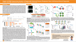 科学海报Single-Cell RNA Sequencing Analysis of Regionally Patterned Human Pluripotent Stem Cell-Derived Neural Organoids


 沪公网安备31010102008431号