Jhaveri DJ et al. (FEB 2010)
The Journal of neuroscience : the official journal of the Society for Neuroscience 30 7 2795--806
Norepinephrine directly activates adult hippocampal precursors via beta3-adrenergic receptors.
Adult hippocampal neurogenesis is a critical form of cellular plasticity that is greatly influenced by neural activity. Among the neurotransmitters that are widely implicated in regulating this process are serotonin and norepinephrine,levels of which are modulated by stress,depression and clinical antidepressants. However,studies to date have failed to address a direct role for either neurotransmitter in regulating hippocampal precursor activity. Here we show that norepinephrine but not serotonin directly activates self-renewing and multipotent neural precursors,including stem cells,from the hippocampus of adult mice. Mechanistically,we provide evidence that beta(3)-adrenergic receptors,which are preferentially expressed on a Hes5-expressing precursor population in the subgranular zone (SGZ),mediate this norepinephrine-dependent activation. Moreover,intrahippocampal injection of a selective beta(3)-adrenergic receptor agonist in vivo increases the number of proliferating cells in the SGZ. Similarly,systemic injection of the beta-adrenergic receptor agonist isoproterenol not only results in enhancement of proliferation in the SGZ but also leads to an increase in the percentage of nestin/glial fibrillary acidic protein double-positive neural precursors in vivo. Finally,using a novel ex vivo slice-sphere" assay that maintains an intact neurogenic niche�
View Publication
产品号#:
05700
05701
05702
05771
产品名:
NeuroCult™ 基础培养基(小鼠&大鼠)
NeuroCult™ 扩增添加物 (小鼠&大鼠)
NeuroCult™ 扩增试剂盒 (小鼠&大鼠)
Katori S et al. (JUL 2009)
The Journal of neuroscience : the official journal of the Society for Neuroscience 29 29 9137--47
Protocadherin-alpha family is required for serotonergic projections to appropriately innervate target brain areas.
Serotonergic axons from the raphe nuclei in the brainstem project to every region of the brain,where they make connections through their extensive terminal arborizations. This serotonergic innervation contributes to various normal behaviors and psychiatric disorders. The protocadherin-alpha (Pcdha) family of clustered protocadherins consists of 14 cadherin-related molecules generated from a single gene cluster. We found that the Pcdhas were strongly expressed in the serotonergic neurons. To elucidate their roles,we examined serotonergic fibers in a mouse mutant (Pcdha(Delta CR/Delta CR)) lacking the Pcdha cytoplasmic region-encoding exons,which are common to the gene cluster. In the first week after birth,the distribution pattern of serotonergic fibers in Pcdha(Delta CR/Delta CR) mice was similar to wild-type,but by 3 weeks of age,when the serotonergic axonal termini complete their arborizations,the distribution of the projections was abnormal. In some target regions,notably the globus pallidus and substantia nigra,the normally even distribution of serotonin axonal terminals was,in the mutants,dense at the periphery of each region,but sparse in the center. In the stratum lacunosum-molecular of the hippocampus,the mutants showed denser serotonergic innervation than in wild-type,and in the dentate gyrus of the hippocampus and the caudate-putamen,the innervation was sparser. Together,the abnormalities suggested that Pcdha proteins are important in the late-stage maturation of serotonergic projections. Further examination of alternatively spliced exons encoding the cytoplasmic tail showed that the A-type (but not the B-type) cytoplasmic tail was essential for the normal development of serotonergic projections.
View Publication

 EasySep™小鼠TIL(CD45)正选试剂盒


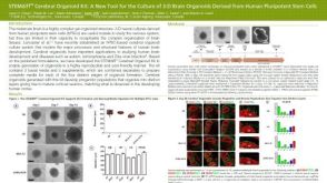 科学海报STEMdiff™ Cerebral Organoid Kit: A New Tool for the Culture of 3D Brain Organoids Derived from hPSCs
 27:19
线上讲座BrainPhys™ Medium Supports the Physiological Activity of Neuronal Tissue in vitro发布日期: 07/22/2016
27:19
线上讲座BrainPhys™ Medium Supports the Physiological Activity of Neuronal Tissue in vitro发布日期: 07/22/2016


 沪公网安备31010102008431号