
 EasySep™小鼠TIL(CD45)正选试剂盒


技术资料
-

-
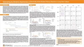 科学海报Determining the Metabolic Requirements of Electrically Active Rodent Primary Neurons in Long-Term Culture
Conference:
Society for Neuroscience Global Connectome 2021
发布日期: 03/17/2021 -
 1:02:03
线上讲座Building Brain Organoids and AssemBloids™ to Study Human Development and Disease发布日期: 10/30/2020
1:02:03
线上讲座Building Brain Organoids and AssemBloids™ to Study Human Development and Disease发布日期: 10/30/2020


 沪公网安备31010102008431号