
 EasySep™小鼠TIL(CD45)正选试剂盒


技术资料
-
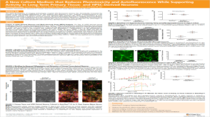 科学海报A New Culture Medium That Reduces Phototoxicity and Autofluorescence While Supporting Activity in Long-Term Primary Tissue- and hPSC-Derived Neurons
Conference:
ISSCR 2019
发布日期: 01/08/2020 -
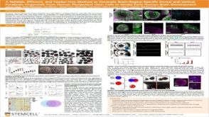 科学海报A Reliable, Efficient, and Feeder-Free Method to Generate Brain-Region-Specific Dorsal and Ventral Forebrain Organoids From Human Pluripotent Stem Cells to Model Early Human Brain Development
Conference:
CSHL 3D Brain 2019
发布日期: 12/20/2019 -
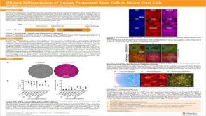 科学海报Efficient Generation of Human Pluripotent Stem Cells to Neural Crest Cells
Conference:
BCRegMed 2019,ISSCR 2019
发布日期: 07/05/2019

 沪公网安备31010102008431号