Utami KH et al. (NOV 2014)
Human mutation 35 11 1311--1320
Impaired development of neural-crest cell-derived organs and intellectual disability caused by MED13L haploinsufficiency.
MED13L is a component subunit of the Mediator complex,an important regulator of transcription that is highly conserved across eukaryotes. Here we report MED13L disruption in a translocation t(12;19) breakpoint of a patient with Pierre-Robin syndrome,moderate intellectual disability (ID),craniofacial anomalies,and muscular defects. The phenotype is similar to previously described patients with MED13L haploinsufficiency. Knockdown of MED13L orthologue in zebrafish,med13b,showed early defective migration of cranial neural crest cells (NCCs) that contributed into cartilage structure deformities in the later stage,recapitulating craniofacial anomalies seen in human patients. Notably,we observed abnormal distribution of developing neurons in different brain regions of med13b morphant embryos,which could be rescued upon introduction of full-length human MED13L mRNA. To compare with mammalian system,we suppressed MED13L expression by short-hairpin RNA in ES-derived human neural progenitors,and differentiated them into neurons. Transcriptome analysis revealed differential expression of components of Wnt and FGF signalling pathways in MED13L-deficient neurons. Our finding provides a novel insight into the mechanism of overlapping phenotypic outcome targeting NCCs derivatives organs in patients with MED13L haploinsufficiency,and emphasizes a clinically recognizable syndromic phenotype in these patients. This article is protected by copyright. All rights reserved.
View Publication
产品号#:
05850
05857
05870
05875
72052
72054
85850
85857
85870
85875
100-1042
产品名:
CHIR99021
CHIR99021
mTeSR™1
mTeSR™1
CHIR99021
Wattanapanitch M et al. (SEP 2014)
PloS one 9 9 e106952
Dual small-molecule targeting of SMAD signaling stimulates human induced pluripotent stem cells toward neural lineages.
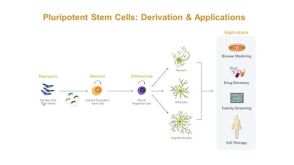
Incurable neurological disorders such as Parkinson's disease (PD),Huntington's disease (HD),and Alzheimer's disease (AD) are very common and can be life-threatening because of their progressive disease symptoms with limited treatment options. To provide an alternative renewable cell source for cell-based transplantation and as study models for neurological diseases,we generated induced pluripotent stem cells (iPSCs) from human dermal fibroblasts (HDFs) and then differentiated them into neural progenitor cells (NPCs) and mature neurons by dual SMAD signaling inhibitors. Reprogramming efficiency was improved by supplementing the histone deacethylase inhibitor,valproic acid (VPA),and inhibitor of p160-Rho associated coiled-coil kinase (ROCK),Y-27632,after retroviral transduction. We obtained a number of iPS colonies that shared similar characteristics with human embryonic stem cells in terms of their morphology,cell surface antigens,pluripotency-associated gene and protein expressions as well as their in vitro and in vivo differentiation potentials. After treatment with Noggin and SB431542,inhibitors of the SMAD signaling pathway,HDF-iPSCs demonstrated rapid and efficient differentiation into neural lineages. Six days after neural induction,neuroepithelial cells (NEPCs) were observed in the adherent monolayer culture,which had the ability to differentiate further into NPCs and neurons,as characterized by their morphology and the expression of neuron-specific transcripts and proteins. We propose that our study may be applied to generate neurological disease patient-specific iPSCs allowing better understanding of disease pathogenesis and drug sensitivity assays.
View Publication

 EasySep™小鼠TIL(CD45)正选试剂盒


 27:19
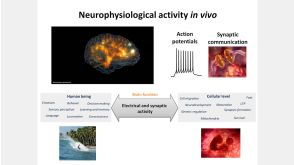
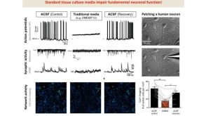
线上讲座BrainPhys™ Medium Supports the Physiological Activity of Neuronal Tissue in vitro发布日期: 07/22/2016
27:19
线上讲座BrainPhys™ Medium Supports the Physiological Activity of Neuronal Tissue in vitro发布日期: 07/22/2016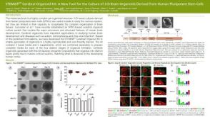 科学海报STEMdiff™ Cerebral Organoid Kit: A New Tool for the Culture of 3D Brain Organoids Derived from hPSCs
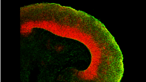
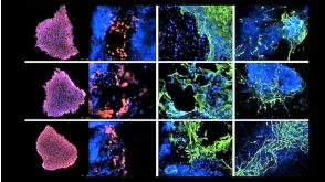
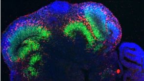

 沪公网安备31010102008431号