
 EasySep™小鼠TIL(CD45)正选试剂盒


技术资料
-
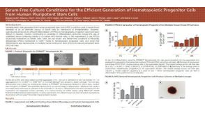
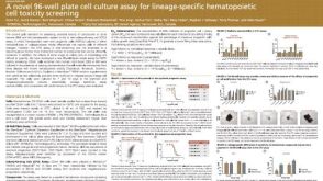 科学海报A Novel 96-well Plate Cell Culture Assay for Lineage-Specific Hematopoietic Cell Toxicity Screening
Conference:
SOT 2015
-
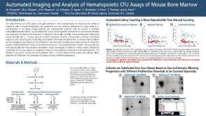 科学海报Automated Imaging and Analysis of Hematopoietic CFU Assays of Mouse Bone Marrow
Conference:
ISEH 2015
-
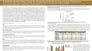 科学海报A Flexible 96-Well Plate Assay for Screening Toxicity to Granulocyte Production
Conference:
SOT 2015
-
 技术窍门为ALDHbr检测分析设置对照和设门

 沪公网安备31010102008431号