
 EasySep™小鼠TIL(CD45)正选试剂盒


技术资料
-

-
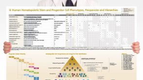 挂图Human Hematopoietic Stem and Progenitor Cell Phenotyping Overview of subset surface markers, frequencies and assays for analysis
 -

-
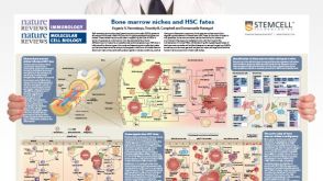 挂图Bone Marrow Niches and HSC Fates A detailed reference on signaling pathways in the bone marrow and how these influence HSC fate decisions; created in partnership with Nature Reviews Immunology and Nature Reviews Molecular Cell Biology
 -
 46:01
线上讲座Targeting Self-Renewal Function in Normal Hematopoietic and Leukemic Stem Cells发布日期: 02/03/2017
46:01
线上讲座Targeting Self-Renewal Function in Normal Hematopoietic and Leukemic Stem Cells发布日期: 02/03/2017

 沪公网安备31010102008431号